Log in    RU


Info
  
Help
  
Images
  
Video
  
Tags


Search 


Main page :: Forum :: Ultrasound cases - Sonograms, cine-loops etc. ::

Hemangioma in the back of the head


Tags: Soft tissues sonography, Images, Video, Clinical report, Esaote MyLab 70, Pediatric

   Posts
 
  Hemangioma in the back of the head
#1

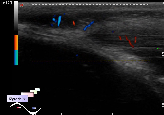
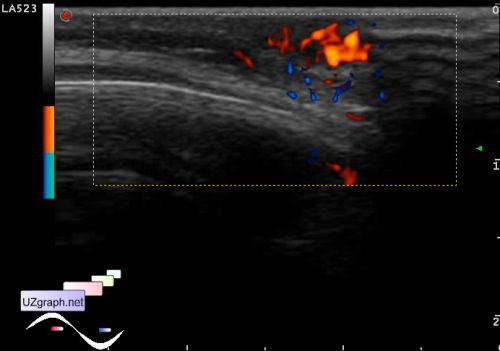
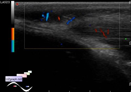
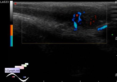
    Child 3 years old with a hemangioma in the occipital region of the head is directed to ultrasound.

    Visually, there is a change in the specified area of the skin, type of birthmark.

    At ultrasound in that area subcutaneously there is a highly blood-supplied hypoechoic oval lesion with an irregular contour, slightly deeper there is a thickened portion of a muscle with increased blood flow at PD and relatively wide vessel (s) up to 1.5 mm (feeding vessel?)

    external link

:: attachments(6) ::

:: file 1 ::

:: file 2 ::

:: file 3 ::

:: file 4 ::

:: file 5 ::

:: file 6 ::