Log in    RU


Info
  
Help
  
Images
  
Video
  
Tags


Search 


Main page :: Forum :: Ultrasound cases - Sonograms, cine-loops etc. ::

Neonatal breast enlargement


Tags: Breast sonography, Soft tissues sonography, Esaote MyLab 70, Images, Video, Clinical report, Pediatric

   Posts
 
  Neonatal breast enlargement
#1

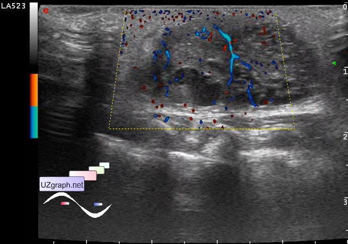
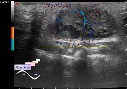
    A male child 1 month old with suspected left breast mastitis.

    Visually left breast enlarged, breast skin is red.

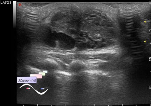
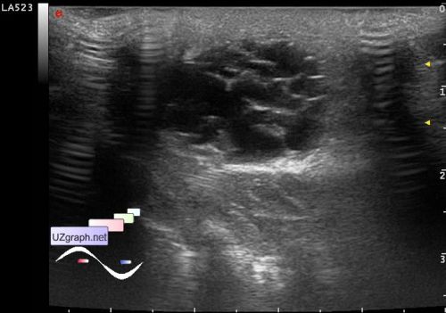
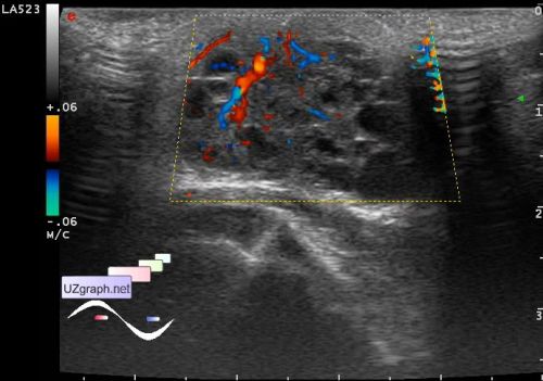
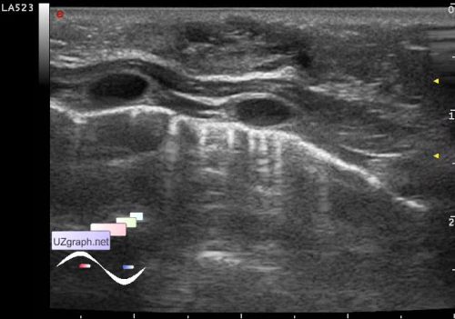
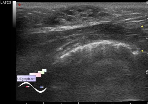
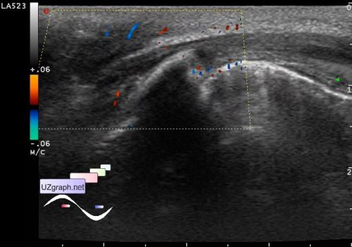
    At US in the projection of both breasts there is an isolated lesions (one on each side) by type of cheese with holes (solid-cystic type), regular oval shape, left size of 2.3 x 1.4 x 2.8 cm, on CFM and DPD richly suppling with blood, right size of 1.9 x 0.6 x 1.8 cm, on CFM with blood flow. In part of the " cystic" zones of the left lesion observed the effect of the movable suspension (?).

    Recommended advice of the oncologist and puncture.

    external link

:: attachments(8) ::

:: file 1 ::

:: file 2 ::

:: file 3 ::

:: file 4 ::

:: file 5 ::

:: file 6 ::

:: file 7 ::

:: file 8 ::

  09:59 02-06-2016
  Case report
#2

    Sonographic features of physiologic neonatal breast enlargement

    external link

  04:06 04-06-2016
  Case report 2
#3

    Sepsis from Neonatal Mastitis and Breast Abscess

    external link

  04:25 04-06-2016
  ref.
#4

    Juvenile papillomatosis of the breast

    external link

    "sometimes termed Swiss cheese disease by pathologists"

    Juvenile papillomatosis and breast carcinoma.

    external link

  14:37 13-09-2019
 
#5

    I found a similar case except that this was a case of a girl - *unfortunately this site no longer exists.*

    Colleagues concluded that this is a telarche, and the cystic component is the ducts.

    So, in my case, it would sound like gynecomastia.