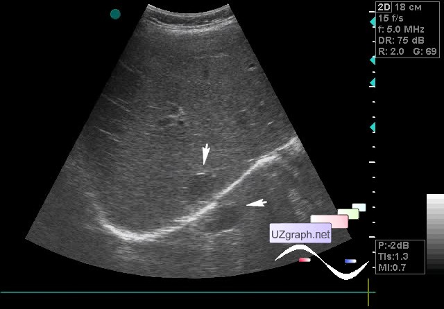
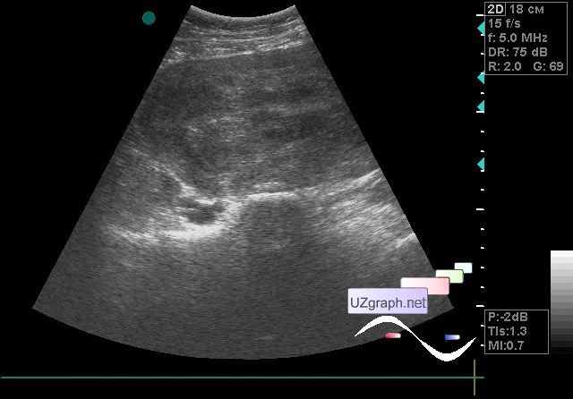
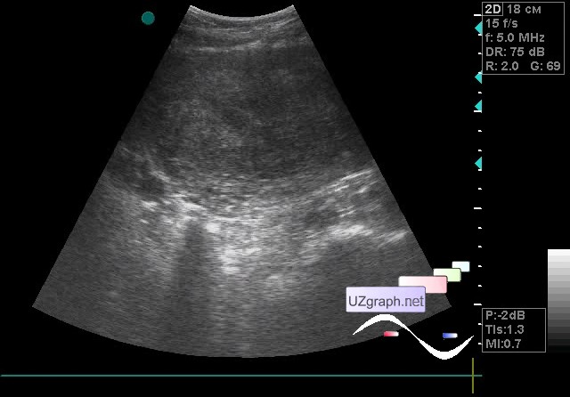
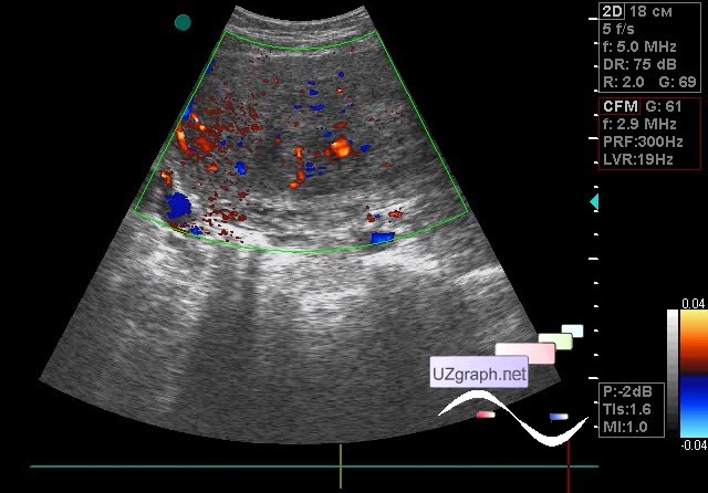
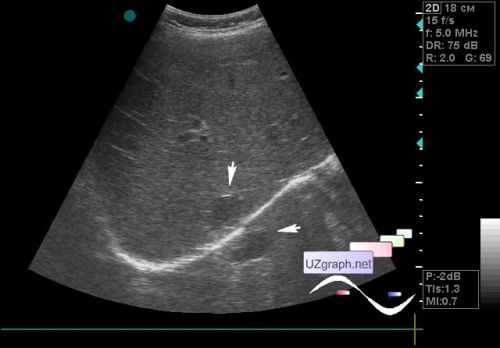
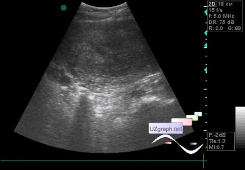
The patient 53 years old came to the abdomen ultrasound, earlier, as far as I understood from her words, there were reported the heterogeneity of the pancreas. On ultrasound the pancreas is normal, in the 7-8 segment of the liver there is a cyst up to 2 cm. At the same time, a mirror artifact was observed (display of phantom cyst upper to the diaphragm). When I tried to put the probe in the pelvis area I felt that I put it on a “stone”, i.e. the abdomen was not compress there at all. The patient said that she had a fibroid/leiomyoma there, with which she had been observed for a long time and that she was saving money for an MRI, and then plans to do an ultrasound ablation of fibroid but hopes that it will pass by itself. At ultrasound the lesion occupying the entire hypogastrium about 15 cm or more in diameter, difficult to say precisely because the mass not completely fit the screen, on the CFM with multiple signals of blood flow (fibroid? other?). Recommended consultation of the surgeon and CT of the abdomen. Ps. Good if it is a fibroid, the main thing if it is not something else - external link external link |