A 5-year-old child from the infectious diseases department of the Children's City Clinical Hospital with a clinical diagnosis: sialodenitis, lymphadenitis. Follow-up ultrasound was prescribed.
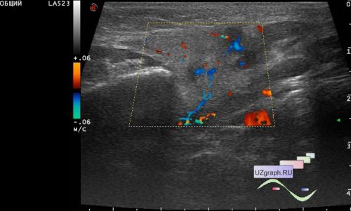
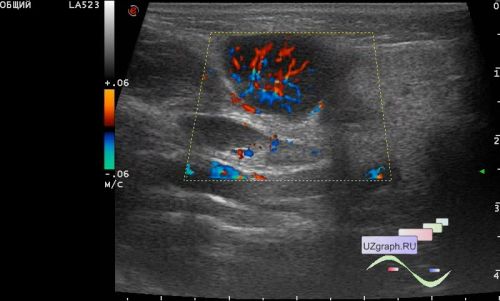
At Ultrasound in the right submandibular region visualizes a hypoechoic lesion of a rounded shape up to 15 x 18 x 11 mm, on the CFM with a pronounced increase in blood flow (differential diagnosis: lymphadenitis, pleomorphic adenoma, lymphadenopathy, etc.), next to another similar up to 15 x 11 mm and another 3-4 pieces. similar up to 9 mm in size, on the CFM with increased blood flow (differential diagnosis: lymphadenitis, lymphadenopathy, etc.).
In the left submandibular region, a single hypoechoic lesion up to 20 x 6 mm is visualized, on the CFM with increased blood flow, a number of similar 5-6 pcs. up to 10mm, on the CFM with increased blood flow. (differential diagnosis: lymphadenitis, lymphadenopathy, etc.).
Also there is an enlargement of the right submandibular salivary gland relative to the left (differential diagnosis: sialadenitis on the right, etc.)