A 2-year-old child, parents went to the public clinic for a follow-up abdominal ultrasound due to a previously identified mass.
According to an extract from one infectious diseases children's Moscow hospital, the child was recently there with an intestinal infection, during the examination, an ultrasound scan revealed a mass near the lower pole of the spleen, as well as diffuse-focal changes of the liver with lesions up to 12 mm. Later, in the same hospital, the child underwent a CT scan of the abdominal cavity, which did not confirm diffuse focal changes in the liver, but only a mass near the spleen, without a more accurate diagnosis, i.e. just lesion.
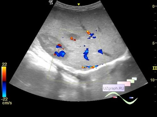
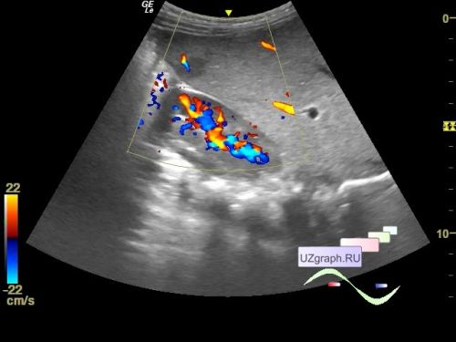
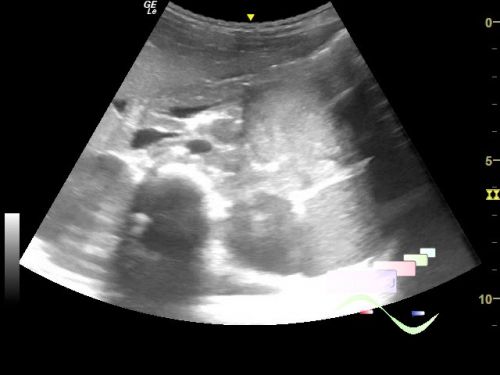
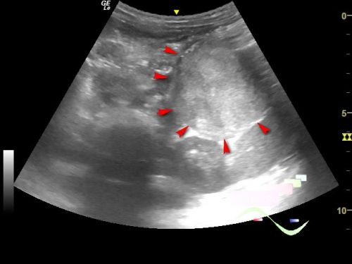
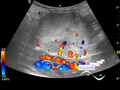
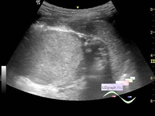
On the current ultrasound, diffuse-focal changes of the liver are visualized, according to the type of hypoechoic rounded inclusions up to 17 mm in size, at CFM with blood flow (differential diagnosis: metastases, etc.), also in the projection of the tail of the pancreas, a heteroechoic lesion is visualized, with blood flow at CFM, up to 5 x 5 x 6.6 cm in size (differential diagnosis: adrenal tumor, etc.).
Recommended: consultation of an oncologist in an oncology center.