A 4-year-old child is observed with a diagnosis of suprahepatic portal hypertension and was referred for ultrasound by a surgeon due to hydroscrotum.
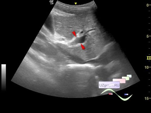
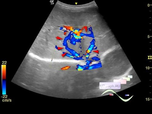
On ultrasound, the portal vein is dilated to 10 mm (norm for age is up to 7 mm), portosystemic collaterals are visualized, the spleen is enlarged to 14 cm (norm for age is up to 10 cm, severe splenomegaly). In the mesogastrium, more than 5 lymph nodes up to 8 mm are visualized (mesadenitis?). Also visualized the cecum with a thickened wall (typhlitis?), a small amount of free fluid with debris in the right iliac region, thickening of the stomach wall (gastritis?), thickening and increased echogenicity of the adjacent omentum (panniculitis?) is noted.
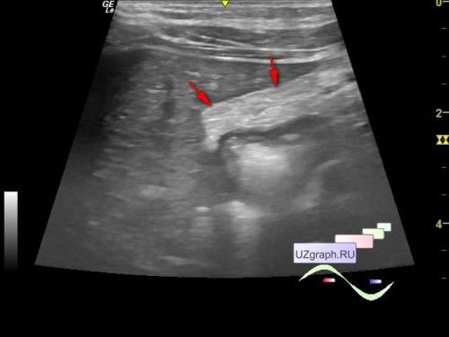
The right inguinal canal in a standing position is dilated, in the right half of the scrotum there is a small amount of free fluid with debris (dropsy/ hydrocele?).