Child 4 years-old with complaints of a painless lesion in the left popliteal area from the surgeon with suspected hygroma aimed to ultrasound of soft tissues of related area.
Visually lesion barely visible only in the standing position, palpable ...
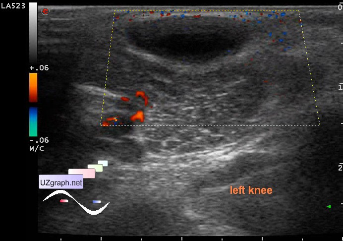
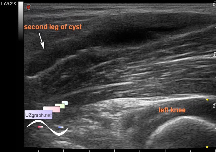
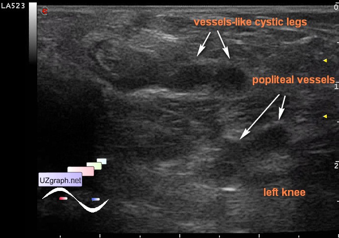
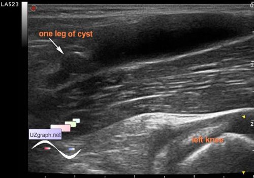
At US in the left popliteal area visualized anechoic lesion, maybe a group of lesions, elongated shape - along the longitudinal axis of the body, coming from popliteal vascular bundle to the skin surface, in the initial section is represented by two parallel tubular structures(vessel-like, "legs" ), size of about 3x1,5cm, without blood flow at CFM(lymphangioma?)
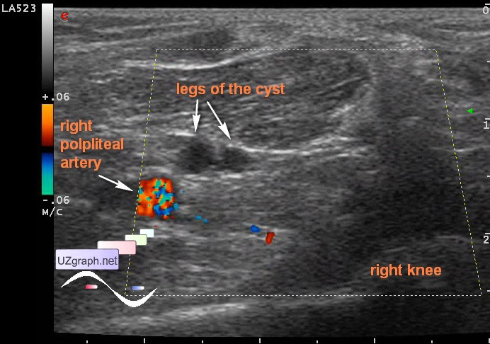
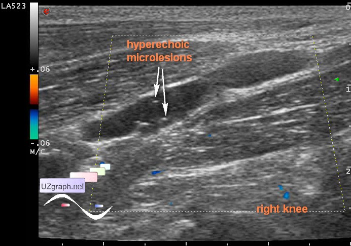
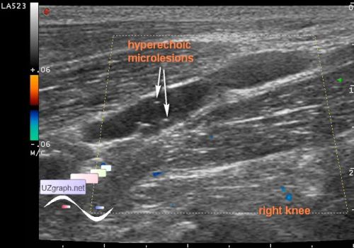
I do not know why, but decided to look for something like that in the right and found almost a twin, just a little smaller in size and with hyperechoic microlesions (microcalcifications? debris? hemorrhage in the cyst?) - bilateral lymphangioma?
"Focal enlargement of veins, arteries, and lymphatic structures around the knee can produce a "cystic" mass. [2],[14] These conditions include aneurysms, varices, hemangiomas, and lymphangiomas.
...
Lymphatic malformations are often present at birth, but may not become evident until later in life. MRI demonstrates a similar appearance to that of venous malformations; they may be macrocystic or microcystic, with the latter form predominating in the extremities"