Log in    RU


Info
  
Help
  
Images
  
Video
  
Tags


Search 


Main page :: Forum :: Ultrasound cases - Sonograms, cine-loops etc. ::

Intestinal obstruction, GB wall thickening


Tags: Abdomen sonography, Gastrointestinal sonography, Images, Video, Clinical report, Esaote MyLab 70, Pediatric

   Posts
 
  Intestinal obstruction, GB wall thickening
#1

    The child 1 month from the pediatrics department, sent to US immediately because of uncontrollable vomiting.

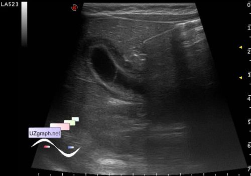
    Gallbladder 30x10mm, the wall is thickened to 4 mm, with increased blood flow at CFM.

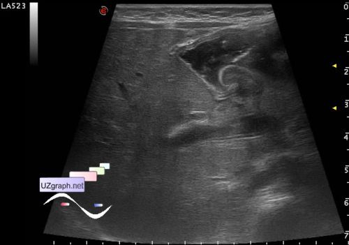
    Stomach, is full, pylorus is retroflexed, pylorus wall to 2.4mm.

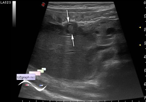
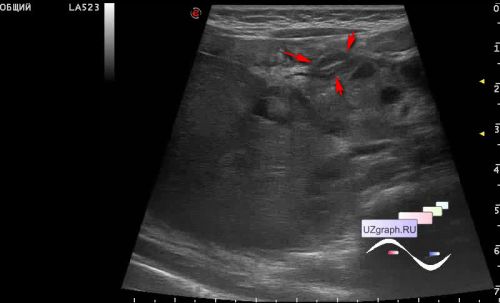
    In the projection in front of the head of the pancreas is visualized lesion about 16x9x12mm, hypoechoic with hyperechoic center part about 5x3x4mm, intimately adherent to the neck of the gallbladder and pylorus (duodenal infiltration? etc.?)

    In most parts of the abdomen mostly empty bowel loops.

    external link

:: attachments(8) ::

:: file 1 ::

:: file 2 ::

:: file 3 ::

:: file 4 ::

:: file 5 ::

:: file 6 ::

:: file 7 ::

:: file 8 ::

  06:36 05-10-2019
 
#2

    Possible differential diagnosis - intestinal malrotation (Ladd' s syndrome).

  17:06 28-04-2024
 
#3

    In the proximal part, an interruption of the duodenum is visualized (video time code 0:30), after all, apparently it was a duodenal atresia.