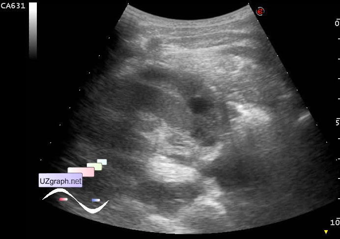
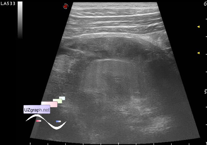
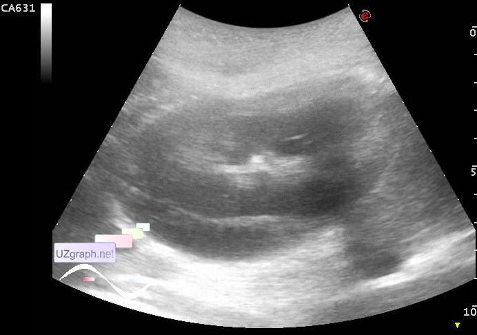
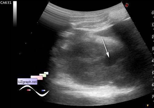
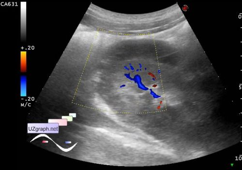
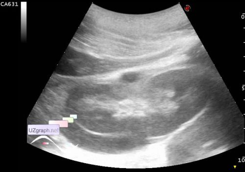
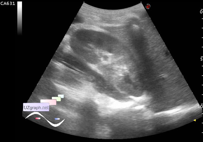
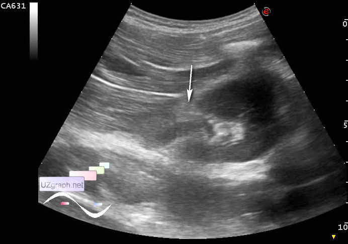
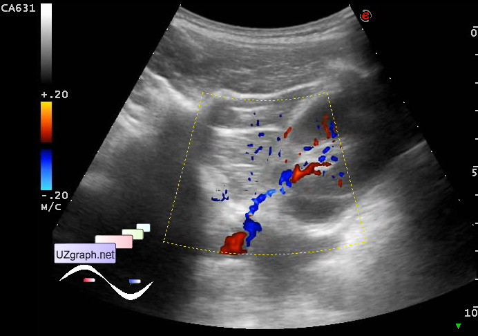
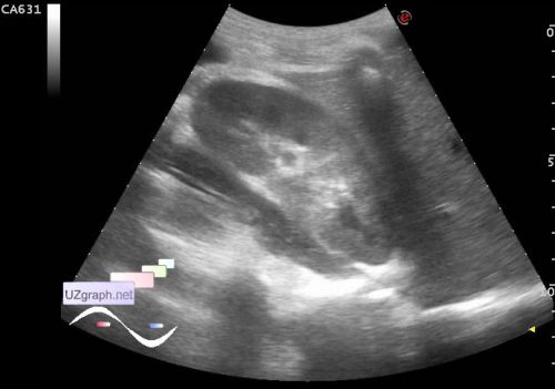
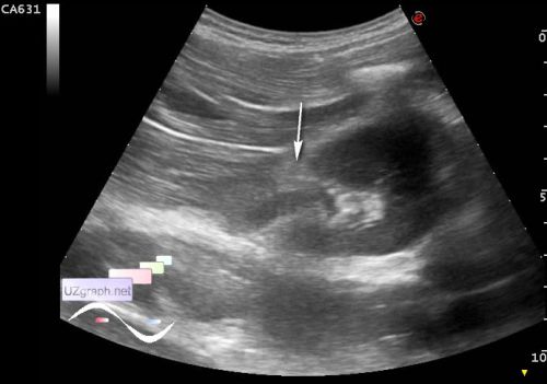
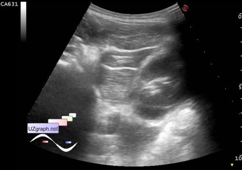
The same child 17 years old with traumatic rupture of the right kidney, renal ultrasound control after 11 days. At the same day (day of first US) child appealed to a some child hospital of Moscow, where it was performed CT which confirmed the right kidney rupture. In the urine analysis red blood cells haven't been identified. Considering that the child refused to comply with bed rest in hospital and there is no need in acute surgical treatment / life-threatening danger at that moment , he was discharged under outpatient monitoring. At the current US, from the side view right kidney looks quite normal, but from the back view: posterior to the kidney perirenal lesion became hyperechoic (organized hematoma?), on the front surface kidney retained a relatively small area with an / hypoechoic content size of about 40x10 mm (fresh hematoma?); PS. Summarizing the interim results, the first ultrasound at 8 days after injury, the second 19 days after injury. And small quote from the hospital paper: "The final clinical diagnosis - kidney injury without open wound in the abdomen. Blunt trauma of the lumbar region. Rupture of the right kidney, perirenal hematoma on the right. ... History of the disease: "got the hit in the right abdomen" - does not specified anything about what kind of hit; the day after the injury - "there were pains in the abdomen, turned to the ambulatory trauma department and was sent to the" - some children hospital of Moscow - "There an acute surgical pathology was excluded. Patient allowed to go home, with the recommendation to do ultrasound of the kidneys ... " - a week later - "made a renal ultrasound in the ambulatory clinic, according to which there was observed accumulation of fluid in the lower pole of the right kidney, child was sent to the " - another children hospital of Moscow - "Gross hematuria didn't found. CT of kidney was achieved without intravenous contrast with CT signs of hematoma of the right kidney. In the urine analysis there was no red blood cell." Well, my conclusion is almost word for word was given in the previous post, the phrase about the "accumulation of fluid in the lower pole of the right kidney" - wasn't in my conclusion, there was described perirenal lesion (rupture of the right kidney? hematoma? urinoma? etc.?), Localization in the lower pole also is not described in my conclusion, because lesion covers more than the lower pole, as someone say, the epistolary genre and "modesty" of some of our clinicians are amazing. There wasn't even a single word about the fact that it was in the ambulatory clinic suspected "the right kidney rupture," and it was written in exact text, and then it turns out that in the ambulatory clinics sitting illiterate doctors because ... according to the rules of fiction, to which apparently can be include a hospital papers, who wrote the "novel" is a "Danko"*.  And guess why most of the hematoma was on the posterior renal contour (gravity in the position of a patient lying on the back), and around the lower pole (gravity in the standing position), as well as why a fresh hematoma at the front contour, in the middle third - in the renal hilum, as can be seen in the CFM (punch in the abdomen). * external link |