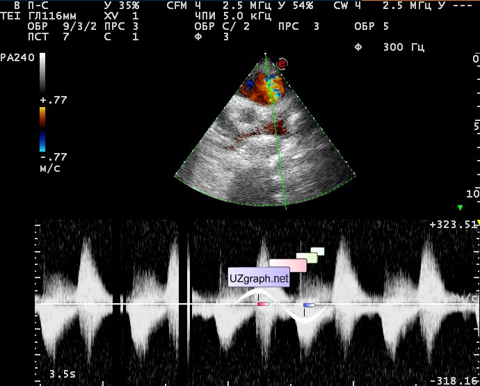
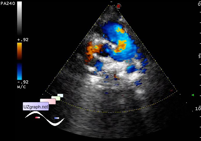
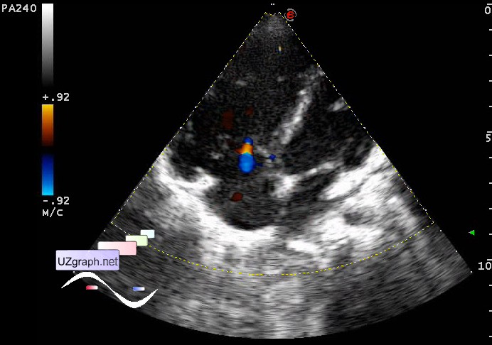
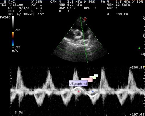
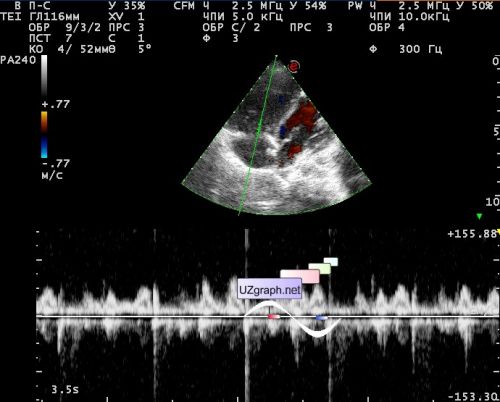
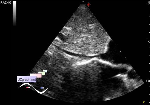
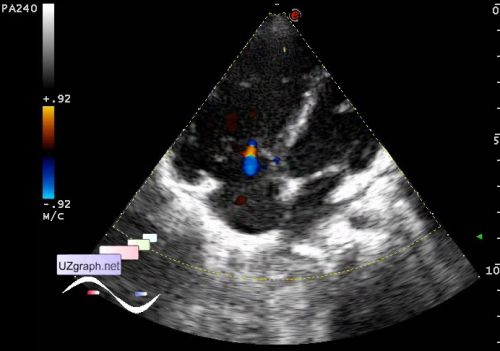
A child of 5 years old from the Ufa town, where she has surgery at 07.11 - Valvuloplasty of PA valve stenosis, PA truncus plasty, PDA ligation. Few quotes from the history: " Fetal ultrasound screening was carried out on time, pathology not revealed ... Echocardiography (06.11): ... ... RV 1,2 cm, RV myocardial hypertrophy ... Pulmonary artery - the diameter of the truncus 1,2 cm ... PA valve - leaflets are thickened, the mobility is limited, MAX PG 74 mm Hg, average PG 39 mmHg Surgery: ... isolated and ligated the PDA (2mm) ... Longitudinal arteriotomy of PA. PA valve is formed as a three-leaflet ... Made valvuloplasty, partial resection of the thickened edges of the leaflets ... Plasty of PA truncus made by auto-pericardium patch. " In October of the same year, examined in Sochi town: " Echocardiogram: RVWT - 4.5mm, the RV - 14 mm, in the trunk of the pulmonary artery systolic blood flow accelerated to 1.8-1.9 m / s (maximum gradient of 14-17 mmHg, the average 7-8 mm Hg) ... Clinical diagnosis: CHD. Condition after surgery .... good result." Next echocardiography data from some Republican cardio-center, from unknown town : " 10.2012 - RV 1.5 cm ... Pulmonary artery moderately enlarged, trunk diameter 1,7cm, pulmonary valve - the fibrous ring of 0.9 cm, leaflets moderately thickened, PG 18 mm Hg ... Pulmonary regurgitation (+ +) . Calculated pressure in the right ventricle is 26 mmHg. Conclusion: Corrected CHD, moderate residual VSPA(valve stenosis of PA). 04.2013 - RV 1.56 cm ... Pulmonary artery - enlarged at the bifurcation, trunk diameter of 1.5 cm, at the bifurcation 2.4 cm ... The pulmonary valve - FR(fibrous ring) - 1,1cm, leaflets moderately thickened, PG 12 mm Hg ... Pulmonary regurgitation (++). Calculated pressure in the right ventricle 26 mm Hg. Conclusion: Corrected CHD ... Moderate residual VSPA. Pulmonary valve regurgitation. 04.2014 - RV 1,9 cm ... Pulmonary artery - is not enlarged, the trunk diameter 1,8 cm, bifurcation 2,4 cm ... Pathological streams in PA is not revealed. The pulmonary valve PG 11 mm Hg ... Pulmonary regurgitation (++) , wide. Calculated pressure in the right ventricle of 23 mmHg. Conclusion: Corrected CHD ... Moderate residual VSPA. Pulmonary valve regurgitation. The increase in size right ventricular cavity. 04.2015 - RV 1.9 cm ... Pulmonary artery - is not enlarged, the trunk diameter of 1.5 cm. Pathological streams in PA is not revealed. The pulmonary valve PG 12 mm Hg ... Pulmonary regurgitation (++), wide. Calculated pressure in the right ventricle is 20 mmHg. Conclusion: Corrected CHD ... Moderate residual VSPA. Pulmonary valve regurgitation. The increase in size right ventricular cavity." Another echocardiogram from 10.2015 without name of medical center, handwritten (something is unreadable): " ... The pulmonary artery FR 12, the truncus 15 mm. Pulmonary valve V 1,6 m / s, R2+, PG PA 10,41 mm hg ... Right ventricle cavity size in diastole 2.35 ... Conclusion. Increased RV ... Fast stream in PA. PR 2+, PG PA 10.41 mmHg... Surgery of CHD. Slight St. PA with PR 2+" . Consultation of cardiosurgeon from some Institute of Moscow, at 10.2015 (possibly last echocardiography from there, again written by hand ....): " There is no data for restenosis. The restrictions do not need." At the current US visualized regurgitation 0-1 st. at TV, Vmax of regurgitation 1.2 m / sec. IVC contraction more than 50% (almost complete). PA diameter (in PV area) 14 mm. In CFM mode regurgitation paints over 100% of PA diameter (as if the valve is not there at all!). Vmax of regurgitation in PA 2.9 m / sec.Vmax of antegrade flow in PA 2 m / sec, PGmax 16 mm Hg .. Also, the retrograde flow is recorded in PA to 2.6 m / sec. PG (RA / RV + RAP) = 11 mm Hg; EDPPA = 39 mm Hg .; AT PA = 133 ms. The curve of the spectrum in PA unchanged throughout the PA. The M-mode registers paradoxical movement of the interventricular septum. RA 32x20mm; EDRVD (apical 4-chamber) 29mm. RVWT 6-7 mm. Conclusion: Stenosis of the PA; Pulmonary hypertension; Overload / dilatation / hypertrophy of the RV. Recommended: urgent consultation of cardiologist; other examination - TE-echocardiography, et al. external link |