Child 4 months underwent several heart surgeries in the cardiocenter, including, as far as I understood, aortic valve replacement and thoracic Ao plastic surgery, with a diagnosis of aortic valvular / supravalvular stenosis (PG 60-80 mmHg), left atrial dilatation, LV endocardial fibroelastosis, low ejection fraction (20-40% Simpson). He's on the waiting list for a transplant. Initially, the parents went to the public clinic with complaints about the child's frequent cough.
He was sent for follow-up echocardiography to the public clinic.
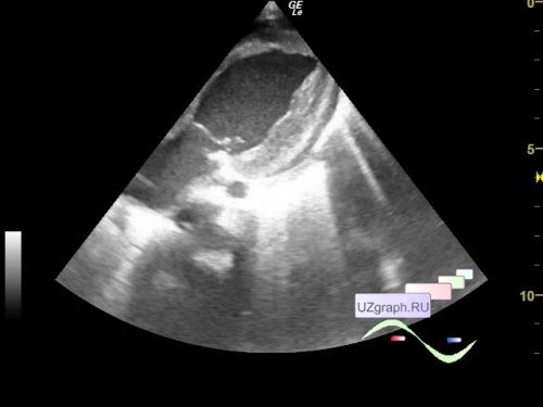
On the current EchoCG, the chambers of the heart are not dilated. Contractility is impaired, hypo-/akinesis of the middle and basal LV segments is observed. LV myocardium with increased echogenicity (differential diagnosis: fibroelastosis, etc.)
With color and spectral Doppler mapping, insufficiencies of 1-2 st. are recorded at mitral, tricuspid, pulmonary valves.
Blood flow velocity in the LVOT up to 1.0 m/s after AV 2.0 m/s (PG 16 mmHg, differential diagnosis: aortic valvular/supravalvular stenosis), pulmonary 1.2 m/s (N<1.1 m/s), in the thoracic Ao up to 2.0 m/sec. Thoracic Ao up to 7 mm, in the projection of the isthmus up to 4.5 mm (differential diagnosis: aortic coarctation, etc.).
The end-diastolic size of the left ventricle is 25 mm, the thickness of the posterior wall of the left ventricle is 13 mm (N<6 mm, myocardial hypertrophy), the ejection fraction is 65 (Teicholz), 38 (Simpson)%.