A 10-year-old child was sent for an ultrasound of the scrotum with a diagnosis of hydrocele, earlier for several years with the same diagnosis was observed in another city.
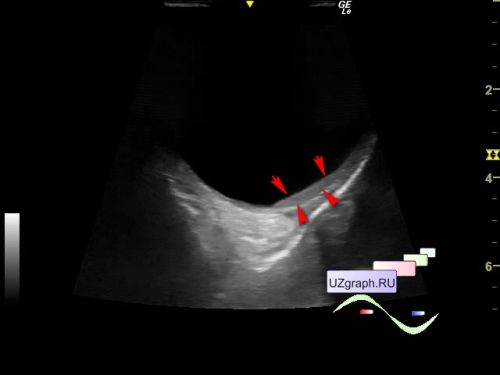
On the current ultrasound in the right half of the scrotum, a lesion type of large cyst, up to 94 x 45 x 45 mm in size is visualized, along the lateral inner wall of the cystic lesion, an echogenic structure is visualized (presumably the right testicle / appendage with deformation / hypoplasia / compression / testicular atrophy), size 24 x 5 x 14 mm.
"One hundred and twenty cases of big unilateral hydrocele of the tunica vaginalis testis have been studied to ascertain the effect on the structure and function of the testis, taking the normal side as control. There was no pressure effect from the hydrocele on the structure of the testis in 70 per cent, a flattening of testis in 22 per cent, and atrophy of testis in 8 per cent of cases. There was partial arrest of spermatogenesis in 10 per cent and total arrest of spermatogenesis in 8 per cent of cases. The remaining 82 per cent showed normal spermatogenesis."