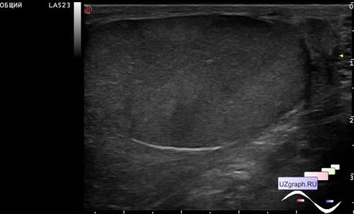
A teenager complaining of an episode of tingling sensation at one-sided of the groin the day before and soreness of the testicle on the same side on palpation turned to the ED of the Children's City Clinical Hospital and was urgently sent for an ultrasound scan with a differential diagnosis - torsion of hydatids, varicocele.
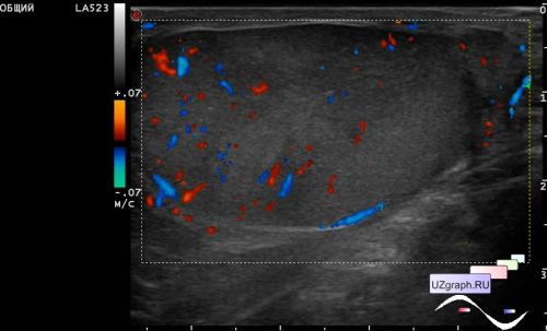
On ultrasound, on the side of complaints, the testicle looks spotty, due to hypoechoic lesions with the echo-phenomenon of "lakes" / "geographical map", on the CFM in the spots, an increase of blood flow is visualized (most likely orchitis).
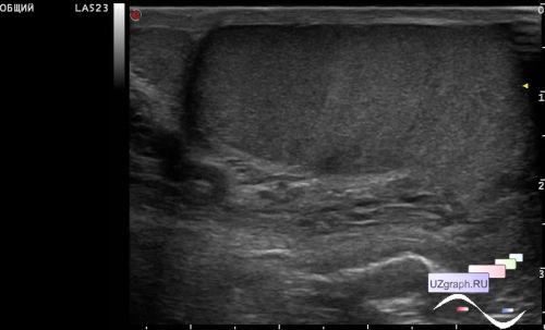
The contralateral testicle also has a diffusely heterogeneous structure like a "puff pie" / "barbell pancakes", due to concentric areas with different echogenicity, also a positive Valsalva test was detected on this side (retrograde venous flow when straining after a deep breath), pampiniform plexus veins up to 3 mm. (congestive orchitis cannot be excluded).