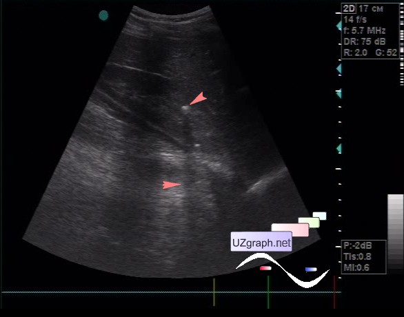
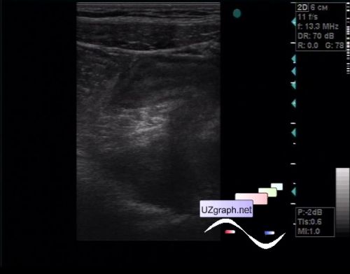
A 36-year-old female patient was referred to an abdomen ultrasound because of pain in hypogastrium. At ultrasound abdomen organs have no specific features. In the right kidney a 5 mm concrement with an acoustic shadow is visualized(unfortunately only the image was saved), the patient told that earlier the ultrasound revealed a calculus in another kidney. In the hypogastrium / small pelvis, the spasmodic part of the intestine is visualized, without peristalsis, without blood flow at CFM, with a thickened upto 6mm wall, passing into the cecum, which at the beginning was regarded as Mekel' s diverticulum, but then I saw that this portion I accepted for the blind end was just a bend on 90 deg. and this is the ileum (ileitis). |