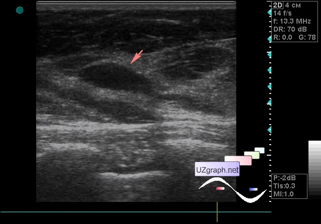
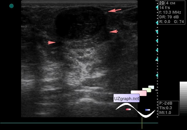
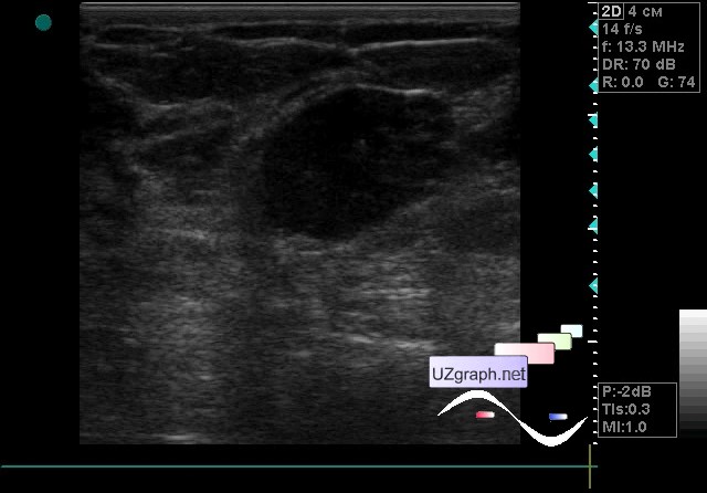
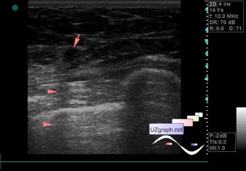
| A 30-year-old patient came to an ultrasound of the mammary gland, according to the patient' s earlier lesions were detected in the left mammary gland. In the right mammary gland, an / hypoechoic lesion of an oval shape, horizontal orientation is visualized, on the CFM without blood flow, up to 1.4 cm in size (cyst? adenoma? other?). In the left mammary gland is visualized 3 lesions: - an / hypoechoic lesion of a teardrop shape, vertical orientation, with an acoustic shadow(possibly as lateral shadows), without blood flow on the CFM, up to 5 mm in size (cyst? etc.?); - hypoechoic lesion of an oval shape, horizontal orientation, on the CFM without blood flow, 2.1 cm in size, with lateral shadows (adenoma? other?); - hypoechoic lesion of a rounded shape, with an uneven contour, horizontal orientation, on the CFM without blood flow, up to 1.7 cm in size, also with lateral shadows (adenoma? else?) external link | |