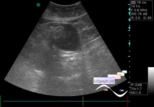
A patient 70 years old came to an ultrasound scan of the abdomen and kidneys with complaints of pain in the back and right flank. At abdomen and kidneys ultrasound without features. In the pelvis to the left of the bladder, a rounded hetero-echogenic, mainly anechoic lesion with an echogenic parietal component, without blood flow at CFM, up to 5.5 cm in size, is visualized (ovary teratoma? cystadenoma? etc.?). PS. The patient said that she had never underwent a pelvis ultrasound scan, therefore a suspicion of teratoma. external link |