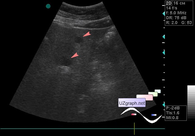
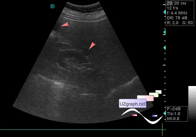
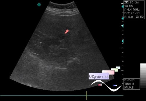
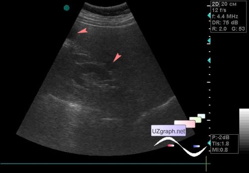
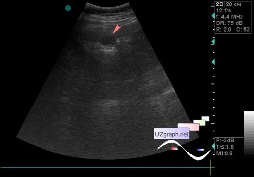
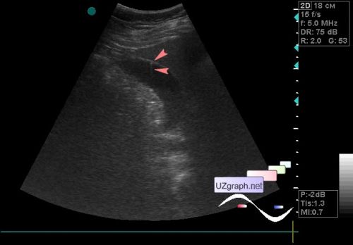
A 68-year-old patient came to an abdominal ultrasound, earlier on an ultrasound cysts were found in the liver and, according to the patient, hemangioma (in the previous conclusion of an ultrasound scan only cysts). At ultrasound in different segments of the liver (2,3,5,6,8) visualizes cysts and 2 hypoechoic zones with a fuzzy contour of up to 2 cm each are visualized near the gallbladder in the projection of 4 segments of the liver (fat-free zones - focal fatty sparing? ). On the front wall of the gallbladder an echogenic inclusion with a comet tail artifact is visualized (cholesterosis - strawberry gallbladder? other?) |