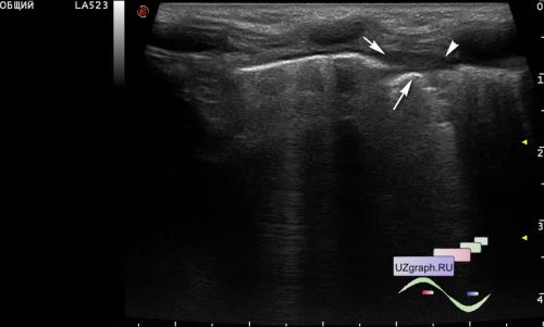
A 1-year-old girl from the infectious diseases department of the Children's City Clinical Hospital with a clinical diagnosis of left-sided pneumonia, an ultrasound of the pleural cavities was prescribed for the amount of free fluid.
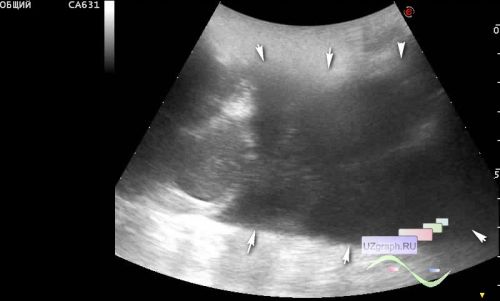
On ultrasound from the anterior approach in the left pleural cavity, the movement of the B-lines is not visualized (hypoventilation), the area of subpleural consolidation is visualized (C-line, differential diagnosis: pneumonia, etc.), from the lateral approach in the left pleural cavity, an airless area is visualized (hepatisation) lung with hyperechoic linear tree-like inclusions (air bronchogram, differential diagnosis: pneumonia, etc.) and about 30 ml of free fluid with septa (hydrothorax).
At the end of the video, for comparison, the normal right lung.