A 3-year-old child in the public clinic was referred for an ultrasound of the parathyroid glands due to hypercalcemia.
On ultrasound, the thyroid gland has a moderately diffusely heterogeneous structure in the posterior sections of both lobes, more on the right (differential diagnosis: iodine deficiency, thyroiditis, etc.)
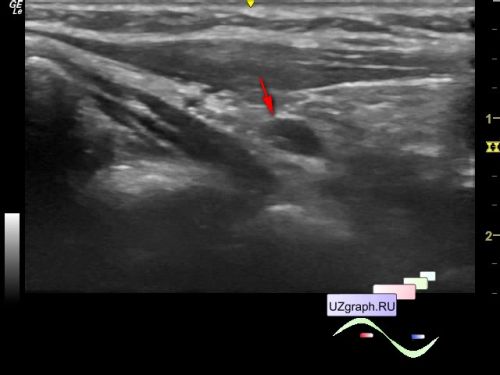
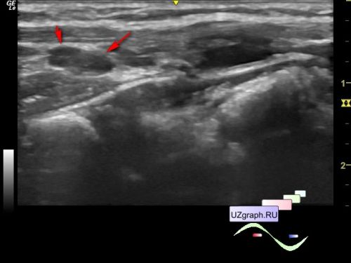
In the projection of the posterior parts of the thyroid gland: on the right, 2 hypoechoic oval-shaped lesions 6x3mm and 4x2mm in size are visualized, the blood flow is visualized on the color flow mode in a larger one; on the left, a hypoechoic lesion of an oval shape up to 8x3 mm in size is visualized, on the CFM with the blood flow (differential diagnosis: adenomas of the parathyroid glands, etc.).