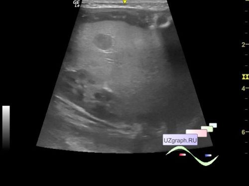
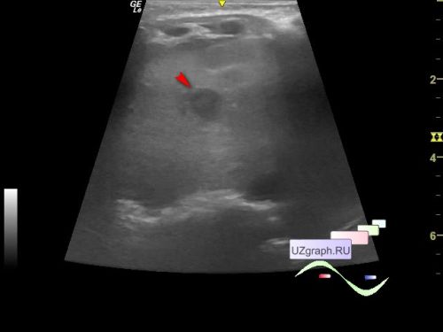
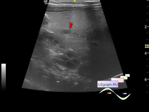
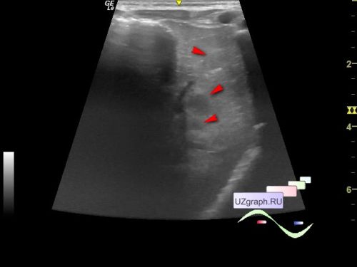
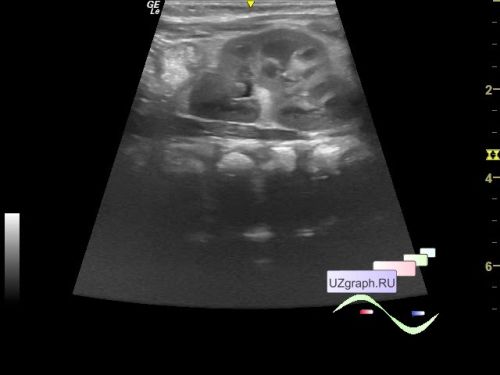
Child 1 month old at the planned screening ultrasound in the public clinic.
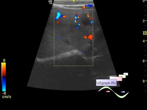
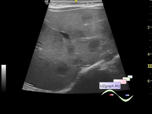
On ultrasound, the echostructure of the liver is diffusely heterogeneous due to hypo- / isoechogenic inclusions of a rounded shape up to 8 mm; it is not possible to assess the blood flow on the CFM, because the child is constantly moving, screaming (differential diagnosis: infantile hemangiomas, abscesses, mts, etc.), the left kidney of a normal structure, size 41x21 mm, ratio 2:1 - thick (differential diagnosis: anomaly of the shape - dromedary hump kidney, nephritis, etc.)
Urgent consultations are recommended: an infectious disease specialist, an oncologist.