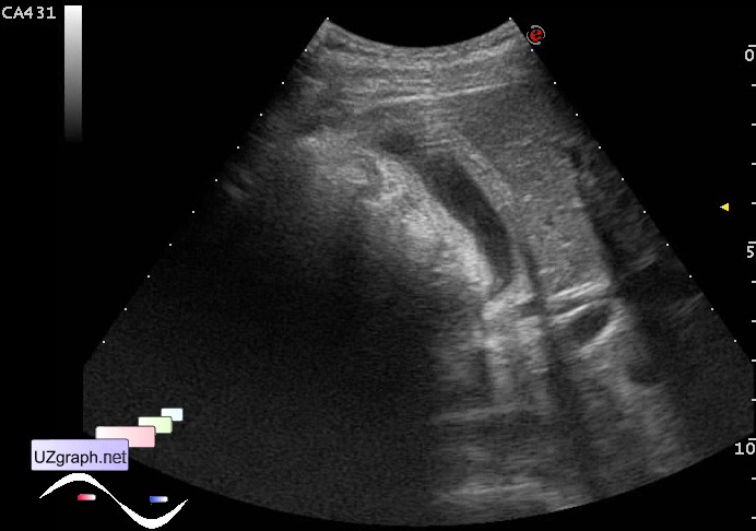
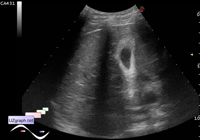
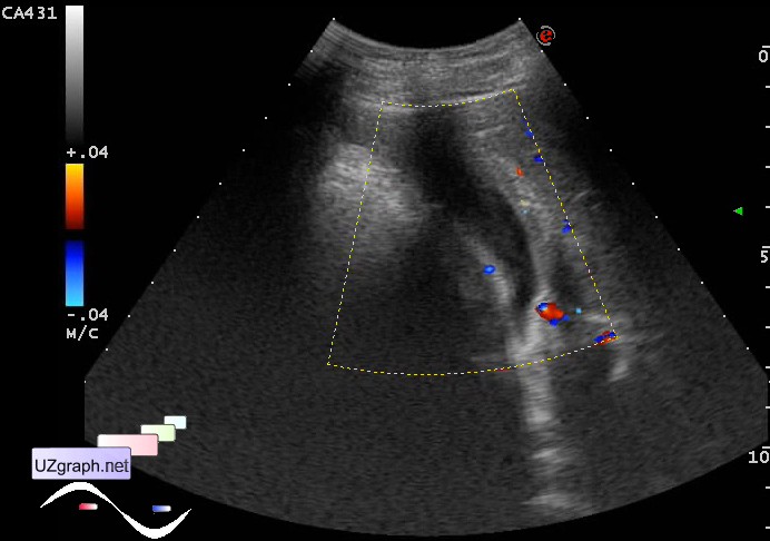
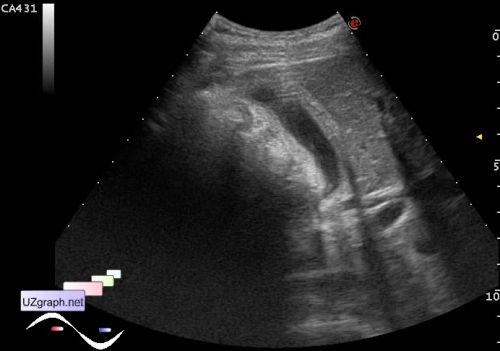
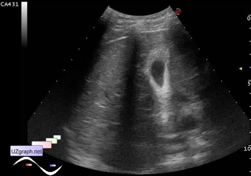
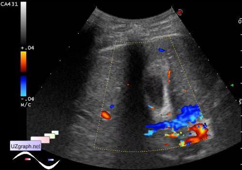
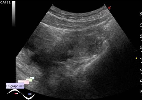
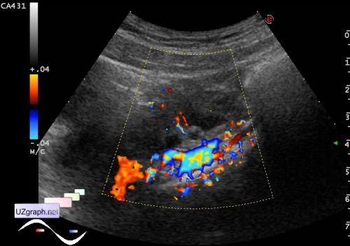
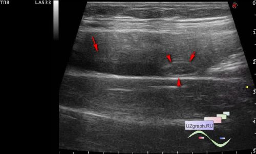
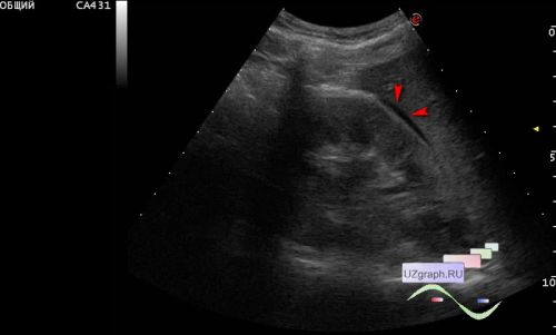
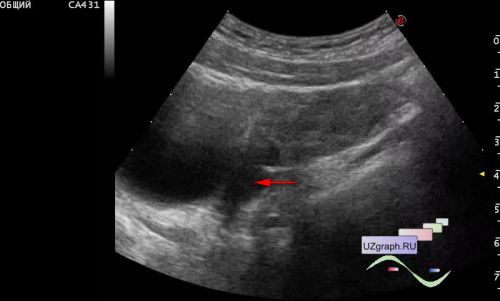
| A teenage girl with complaints of pain in the right hypochondrium was delivered by the ambulance team to the emergency surgical department of the Children's City Clinical Hospital, and was urgently sent for an ultrasound scan with suspected appendicitis. On ultrasound, in the projection of complaints of pain, the gallbladder is visualized with a wall thickening of about 6 mm, at CFM with increased blood flow in the wall (differential diagnosis: cholecystitis, etc.); the appendix is not enlarged(up to 5 mm), in the right parts of the small pelvis visualized a right ovary about 5-6 cm long, at CFM with blood flow (differential diagnosis: oophoritis of the right ovary, torsion of the right ovary, etc.) In the Morison's Pouch (hepatorenal space), in the right lower quadrant of the abdomen (up to 10 ml) and in the small pelvis under the right ovary a small amount of free fluid is visualized.
| |