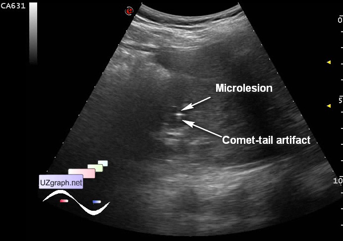
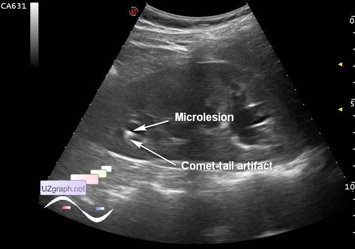
| The patient in the 3 trimester of pregnancy began to disturb back pain, have been made several ultrasound of the kidneys, at the first ultrasound was detected microurolithiasis (1 stone in the top cup of one kidney), on the other ultrasound stones have not been identified, aimed at third ultrasound because pain periodically continue. At US visualized dilatation of calices of corresponding kidney, more pronounced in the upper third, in the lower and middle thirds there is a microlesions with comet tail artifact, 7 mm and 5 mm respectively (microstones?). PS. Despite the fact that small stones must be more visible in the prone position, because kidney located closer to the probe, less noise and so on, nevertheless smaller calculus was detected from supine oblique position(side-view). external link | |