Log in    RU


Info
  
Help
  
Images
  
Video
  
Tags


Search 


Main page :: Forum :: Ultrasound cases - Sonograms, cine-loops etc. ::

Condition after RC PAVC


Tags: Cardiac sonography(Echocardiography), Esaote MyLab 70, Images, Video, Clinical report, Pediatric

   Posts
 
  Condition after RC PAVC
#1

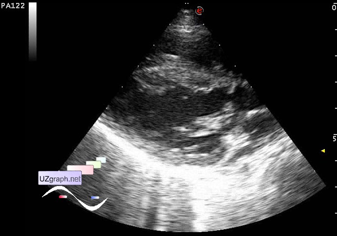
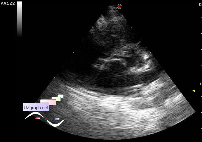
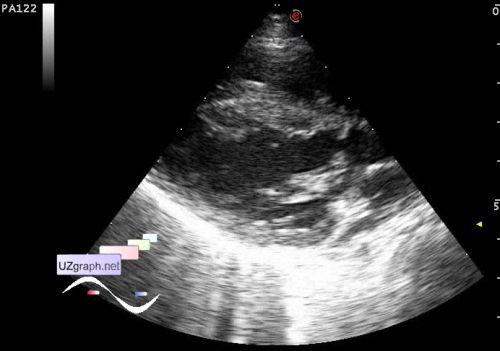
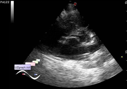
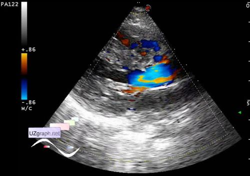
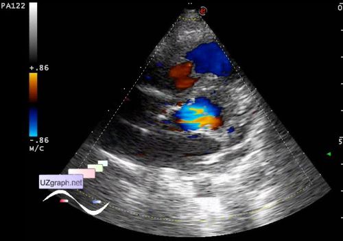
    Child 2 years-old condition after radical correction of partially open atrioventricular canal in 2013. According to relatives, after surgery revealed the membrane in LVOT with PGmax to 35 mm Hg. Under control of a cardiologist in some childrens hospital of Moscow with recommendations of massage, swimming and limit phys. load ...

    Came for the abdominal & kidney US - without features. At the same time I' ve done a quick sighting look on LVOT (a restless child).

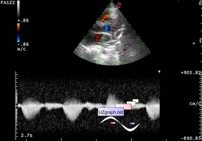
    In the projection of LVOT visualized accessory structure type of the second valve, gradient to 64 mmHg (Vmax 4 m/s)

    external link

:: attachments(6) ::

:: file 1 ::

:: file 2 ::

:: file 3 ::

:: file 4 ::

:: file 5 ::

:: file 6 ::

  09:55 05-10-2019
 
#2

    A colleague from one medical social network said that such membranes in LVOT are called discrete subaortic stenosis.

    external link