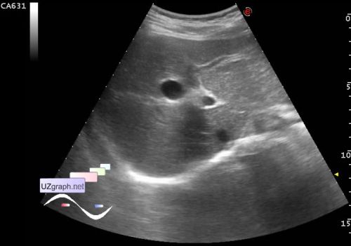
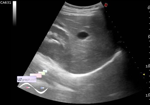
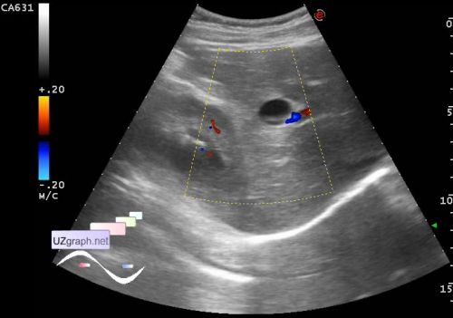
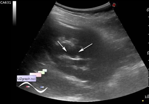
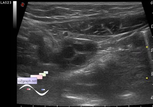
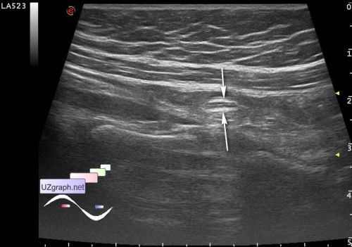
A child 10 years old with suspected appendicitis aimed at US, warned that how she understood that she has a small gall bladder and fused two kidneys. On US Gall bladder is not visualized in the projection of the gallbladder is visualized anechoic round lesion type like cyst, no blood flow at CFM (gallbladder agenesis, choledochal cysts?). The right kidney with parenchymal bridge (duplex? Fig. 4); Calyces to 5.5mm, in the lumen hyperechoic microinclusions to 1 mm (microlites? Fig. 5). The left kidney is not visualized. In the right iliac region is visualized the ovary(Fig. 6) and normal appendix (Fig. 7). external link | 

