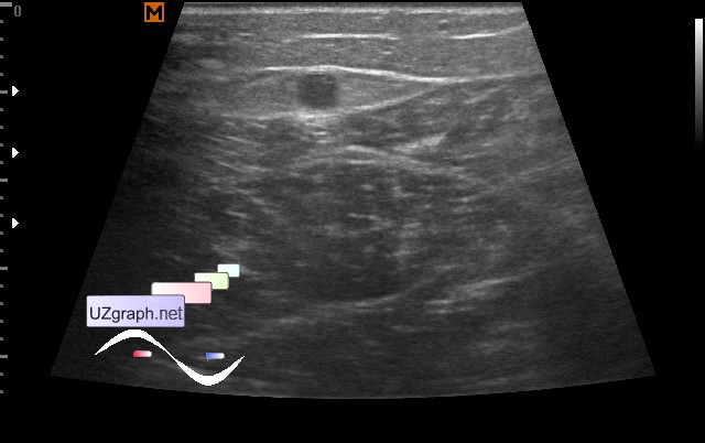
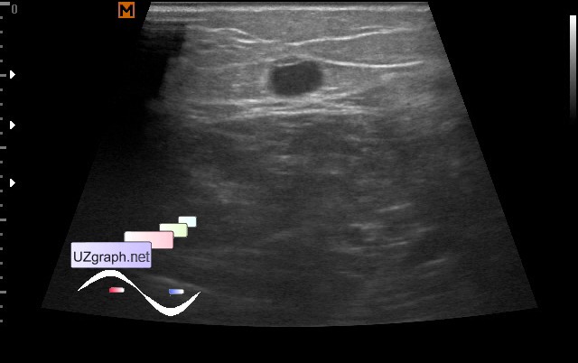
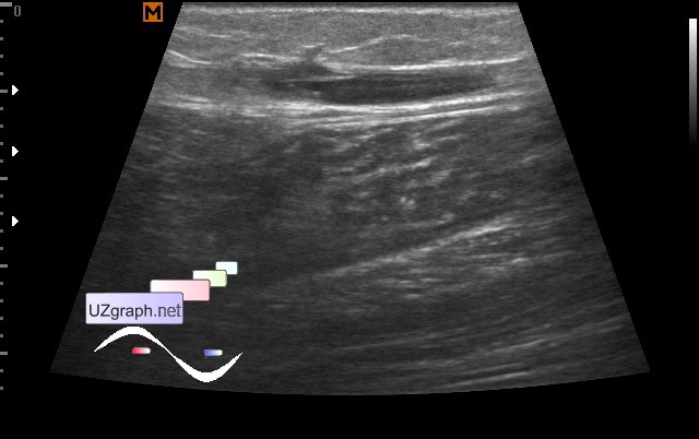
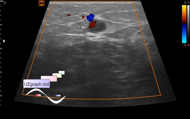
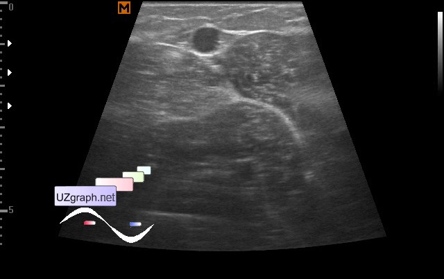
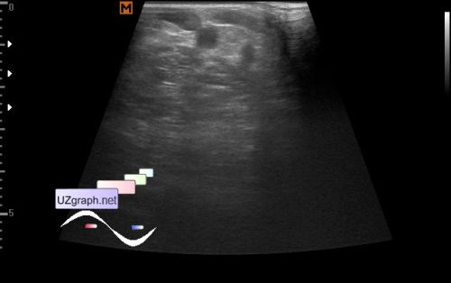
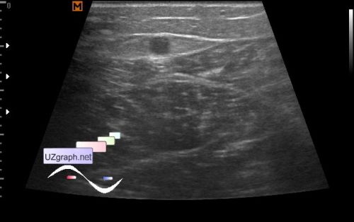
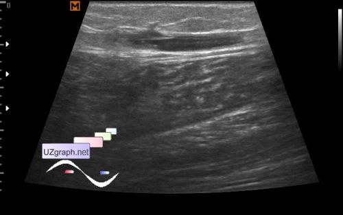
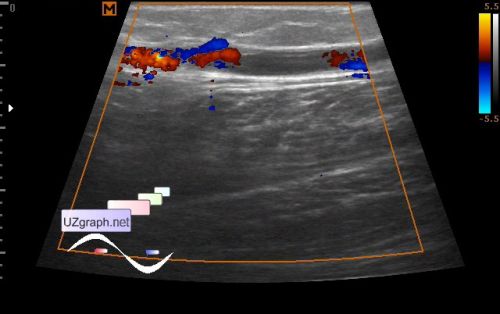
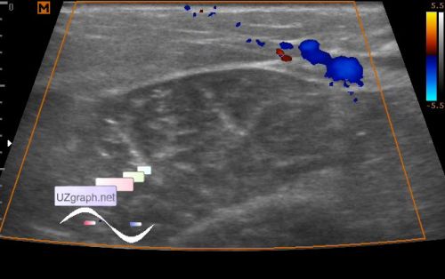
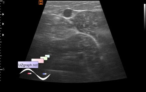
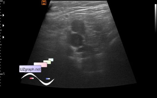
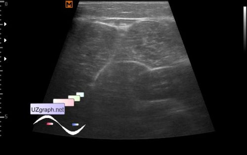
| A girl of 17, a sportswoman, felt pain in the shin during training, turned to the surgeon, a varicose disease was diagnosed at father, a shin injury in the anamnesis is denied. The surgeon identified the strands along the medial surface of the shin, with suspicion of varicose veins sent the child to the US of veins. On ultrasound on the medial surface of the right shin, visualized up to 4-5 mm multiple dilated veins of the GSV system (Great saphenous vein, v. saphena magna, fig.1) from one of them to the surface (in the image upwards) goes the communicating-perforating vein(fig. 4-7) which is lost in the posterior surface of the shin near to the dilated SSV (Small saphenous vein, v. saphena parva, fig.8), near to the point of entry of communicating-perforating vein into the GSV, an open valve of the GSV is visible(fig. 3-6), a transverse section gives an impression of a possible subvalvular thrombosis(fig.3). The SSV is expanded to 5 mm up to the confluence(fig.9) with the popliteal vein. In the standing position the diameter of the described veins increases. Hip veins are not dilated. PS. A sign of the " Egyptian eye" formed by the GSV(fig. 2-3) or SSV(fig.8) and fascias is well visible. For comparison, at the end of the video, a normal " Egyptian eye" is shown from the contralateral lower leg(fig. 10). external link | |