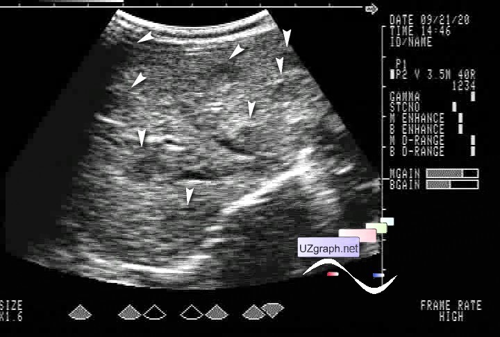
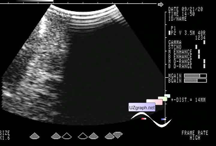
Case from 2007 year. The one-month-old child was sent for an abdominal ultrasound to the public clinic for a routine examination before vaccinations. From the anamnesis, it is only known that the child is undergoing treatment for unilateral congenital dislocation of the hip in one of the research institutes. Otherwise, according to the pediatrician, child is clinically healthy. On ultrasound, the liver is of a diffusely heterogeneous structure due to various-sized hypoechoic lesions. (An ultrasound machine without a Doppler!) Similar spleen lesions have also been suspected. (Later were not confirmed, and upon detailed examination of the cine loops, it is highly likely that they are lesions of the left lobe of the liver, which was visualized through the left lateral approach) Despite the fact that the child went through 2 hospitals and oncological Institute, the final diagnosis of liver lesions was never made, the treatment was not carried out, because the child's condition was assessed as satisfactory. By the year of life, all lesions disappeared on their own. Dif. diagnosis: multifocal form of infantile liver hemangioma, etc. external link |