A 16-year-old teenager from the infectious diseases department of the Children's City Clinical Hospital with suspected appendicitis was sent for an ultrasound scan.
On ultrasound in the right upper quadrant of the abdomen, a section of the intestine with the thickened wall up to 5 mm is visualized (differential diagnosis: enterocolitis, etc.)
In the mesogastrium, more than 10 lymph nodes up to 12 mm are visualized (differential diagnosis: mesentery, etc.)
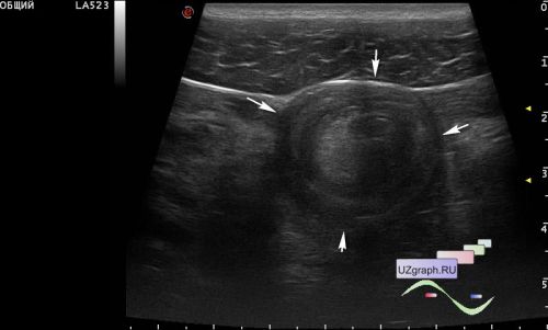
The appendix is not visualized, but in the hypogastrium centrally the target sign is visualized(intestinal intussusception).
On the follow-up ultrasound a few hours later after the enema, intussusception is no longer visualized.