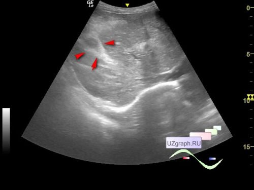
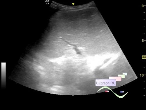
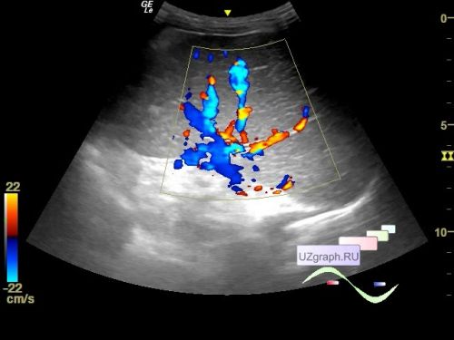
A 3-year-old child was referred by a surgeon for a scrotum ultrasound with suspected scrotum hydrocele and for a follow-up abdominal ultrasound. Previously, the child was examined in one of the Federal Children's Hospitals due to abnormalities in blood biochemistry and ultrasound revealed: cavernous transformation of the portal vein, increased echogenicity in the area of portal vein, portal hypertension, liver enlargement due to the left lobe, splenomegaly and diffuse changes of the pancreas.
On the current ultrasound, as it was also noted in the ultrasound protocol from the children's hospital, he is restless, constantly moving and screaming, which makes visualization difficult, there is indeed moderate scrotum hydrocele, the liver is slightly enlarged, has moderately diffusely heterogeneous structure, the portal vein is cavernously transformed, there is an increase echogenicity around the portal vein, (differential diagnosis: periportal fibrosis, etc.), the gallbladder wall is slightly thickened, the portal vein is up to 3 mm, the spleen is up to 14 cm in maximum size (+4 cm from the normal), the splenic vein is up to 6 mm (normal up to 5.4 mm, differential diagnosis: portal hypertension, etc.), pancreas without features.