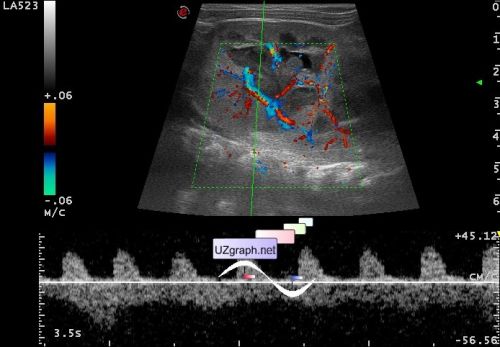
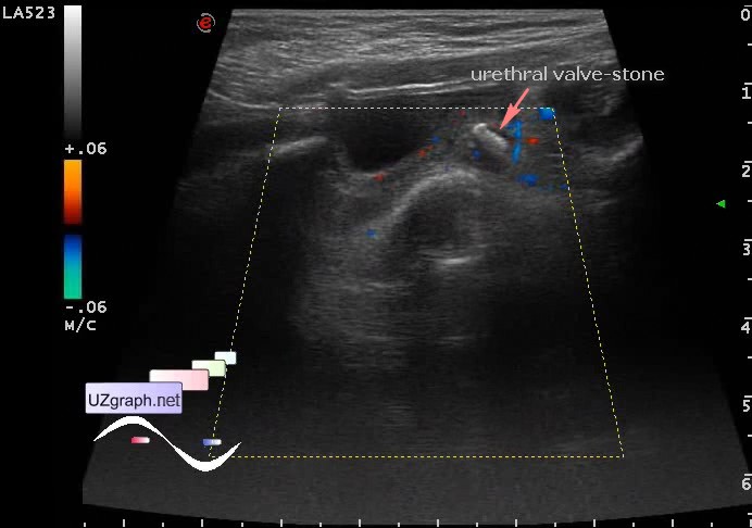
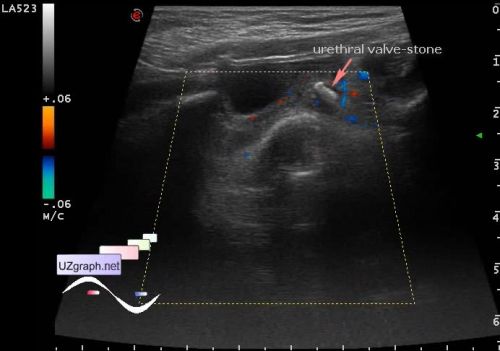
| Baby 2 month old from the cardiology department aims to control ultrasound with suspected abdominal mass, previously diagnosed the blockage(obstruction) of the right kidney. At US kidneys: right 5x3 cm, parenchyma 1.5 cm, left 6,6x3,6 cm (exceeding normal ratio length/ width - 2:1.1 - nephritis?), parenchyma 1.9 cm; corticomedullary differentiation saved. Urinary tract: in the right is not dilated, in the left calyces to 7 mm, pelvis up to 10mm - with hypoechoic content (sludge? pus? etc.?). Right ureter is not visualized, in the left distal part of ureter in the area of bladder inlet visualized hyperechoic irregular shape lesion, size of 3x3x6 mm, without acoustic shadow, without blood flow at CFM (calculus?). Right RI 0.7; Left 0.9 (increased) The bladder is moderately filled, without features. external link | |