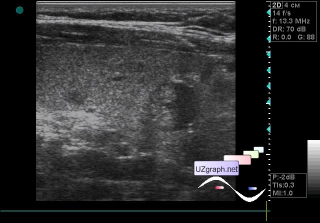
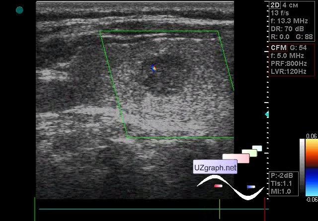
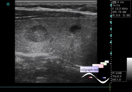
| A 60-year-old patient came to control an ultrasound of the thyroid gland with a diagnosis of a multinodular goiter. In the previous conclusion of the ultrasound examination several lesions were described, including the lesion of calcinate type. On ultrasound in the right lobe about 6 polymorphic lesions are visualized: 1) heteroechoic (hypo / iso), oval shaped up to 1 cm in size, at CFM with the blood flow; 2) up to 5 mm, oval in shape, anechoic with near-wall component, on the CFM without blood flow; 3) up to 3mm, hyperechoic with acoustic shadow(calcinate?); 4) up to 1 cm, oval, anechoic with near-wall component, on the CFM without blood flow; 5-6) hypoechoic oval shape, size 3-4mm. external link | |