
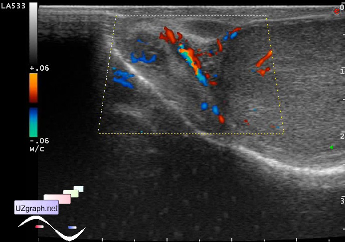
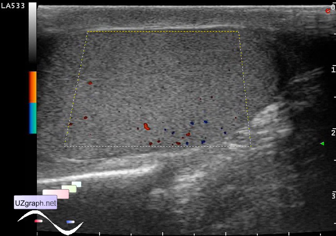
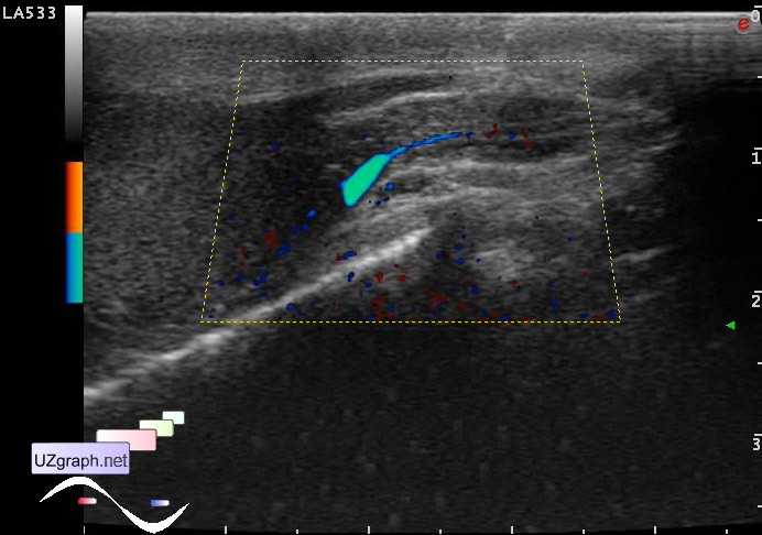
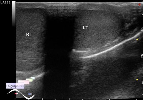
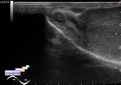
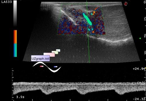
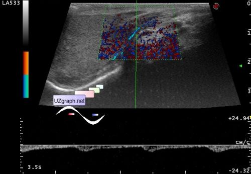
Clinically suspected epididymitis
Tags: Scrotum sonography, Images, Video, Clinical report, Esaote MyLab 70, Pediatric
| Posts | |||
| Clinically suspected epididymitis | #1 |
| |||||
:: file 1 ::
 :: file 2 ::
 :: file 3 ::
 :: file 4 ::
 :: file 5 ::
 :: file 6 ::
 :: file 7 ::
 :: file 8 ::
 :: file 9 ::
 :: file 10 ::
 :: file 11 ::
 :: file 12 :: | |||||


| 18:20 08-05-2022 | #2 |
| |||||