Teenager without complaint with diagnosis of varicocele from prophylactic medical examination by the results of an ultrasound. Urologist did not like this diagnosis and sent for re-ultrasound.
The first thing which immediately have made me distrustful is the resume - " Bilateral varicocele 3 stage" . I do not use the diagnosis of varicocele with stages because the classification of stages is clinical! This alone could cause urologist' s distrustfulness. Also bilateral varicocele is quite rare.
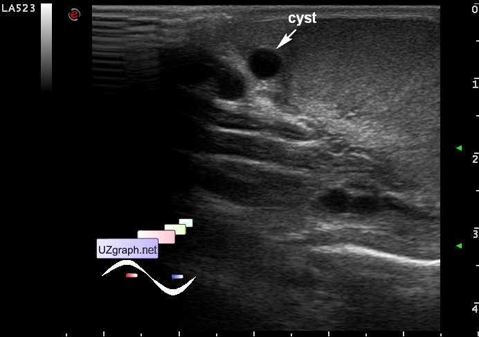
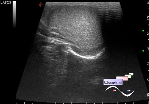
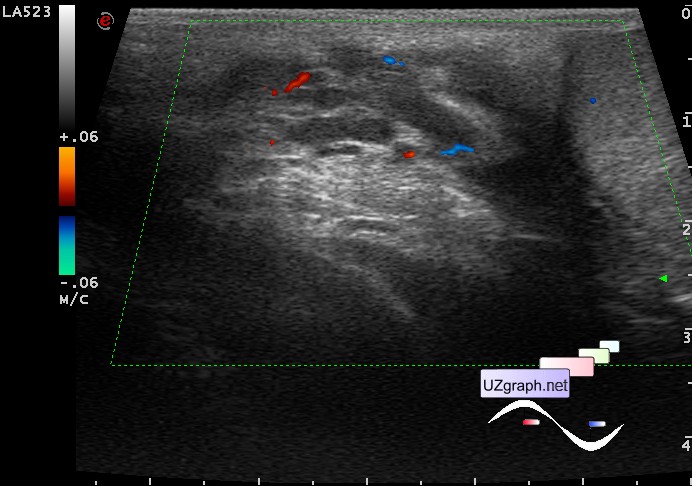
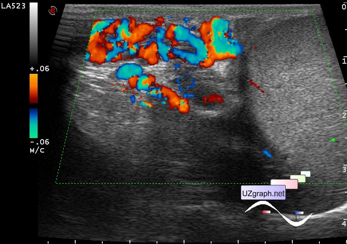
As the result of re-ultrasound, varicocele, as it often happens, only at the left.
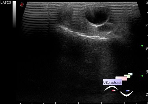
In the spectral doppler long retrograde flow * (* Cornud et al.1999 more than 2 sec.). In the same testicle while the CFM has become clear that there is a cyst / appendage testis / epididymis (arrow).
Returning to the method of evaluation of varicocele in the spectral Doppler mode, another example
Teenager with complaints of recurrent pain in the right testicle, that is very interesting, because everything is allright with it by ultrasound. At ultrasound of the left testicle in CFM Valsalva maneuver doubtful, veins of the pampiniform plexus: resting upto 3 mm, standing upto 4 mm. An attempt of Valsalva maneuver in standing position in PW shows short * retrograde flow (less than 1 second long - physiological by Cornud et al.1999), which explains the dubiousness of the Valsalva maneuver in the CFM, the CFM has always visualized with a delay even technically, and for eyes is hard to catch coloration changing of about 400 msec long.
And what in the end, is there a varicocele or not, how do you think?
Files:
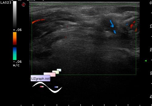
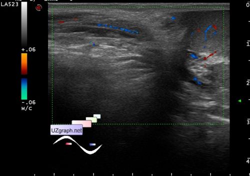
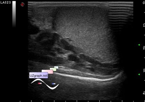
1- normal right testicle
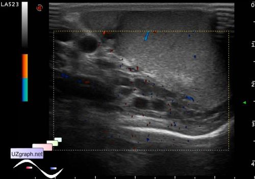
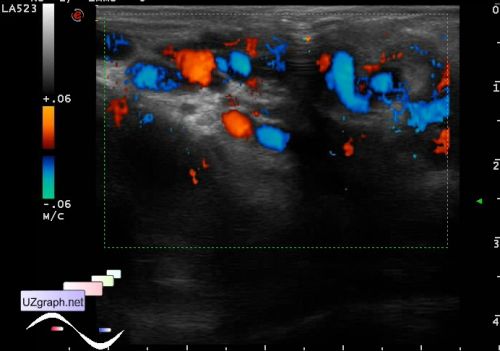
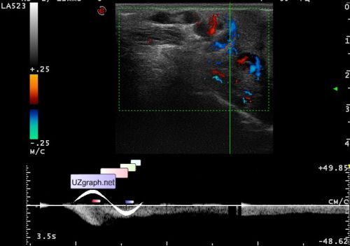
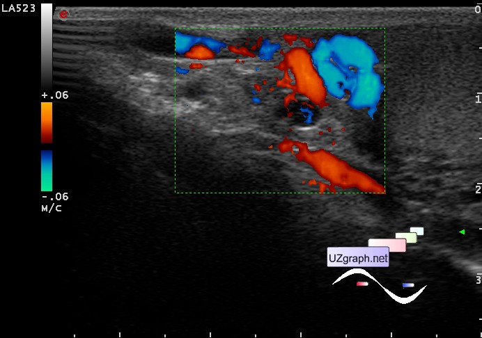
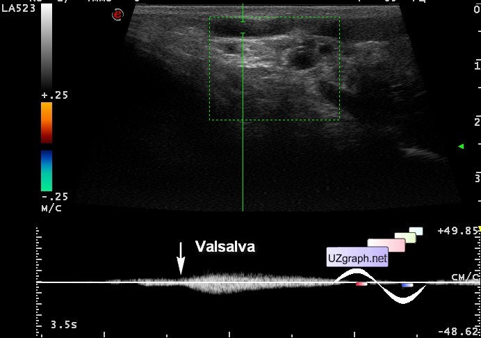
2-4 left testicle:
2 B-mode,
3 CFM standing,
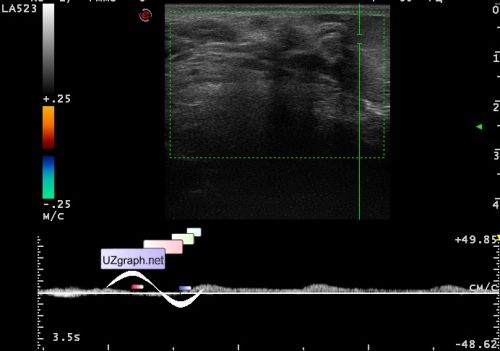
4 PW standing with Valsalva maneuver and corresponding to it a brief inversion of blood flow (below the baseline).
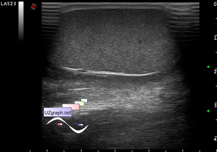
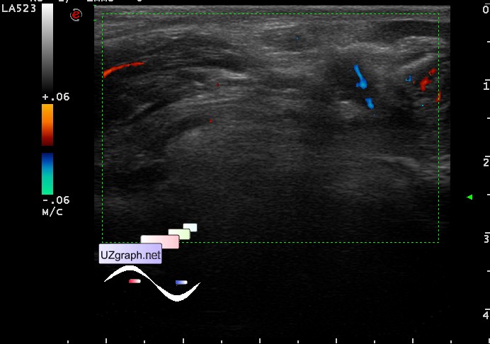
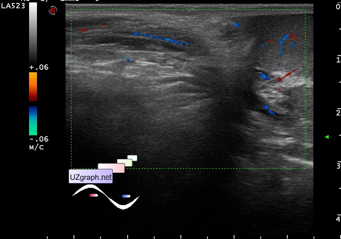
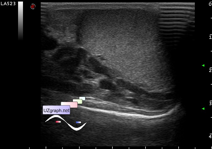
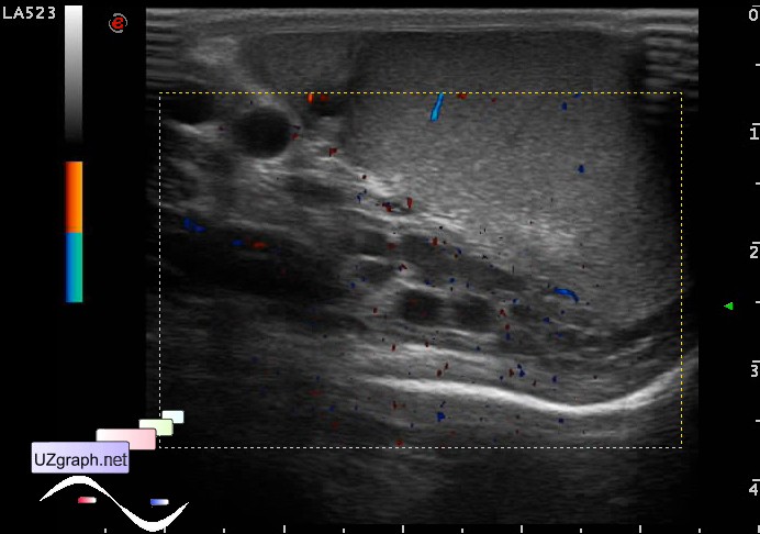
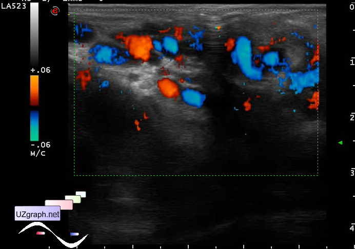
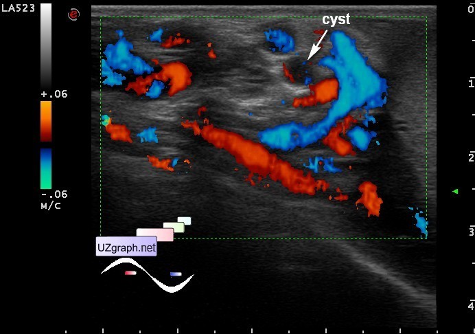
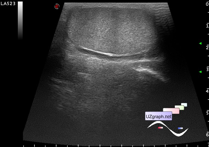
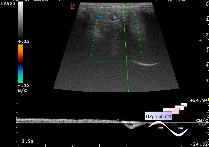
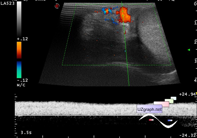
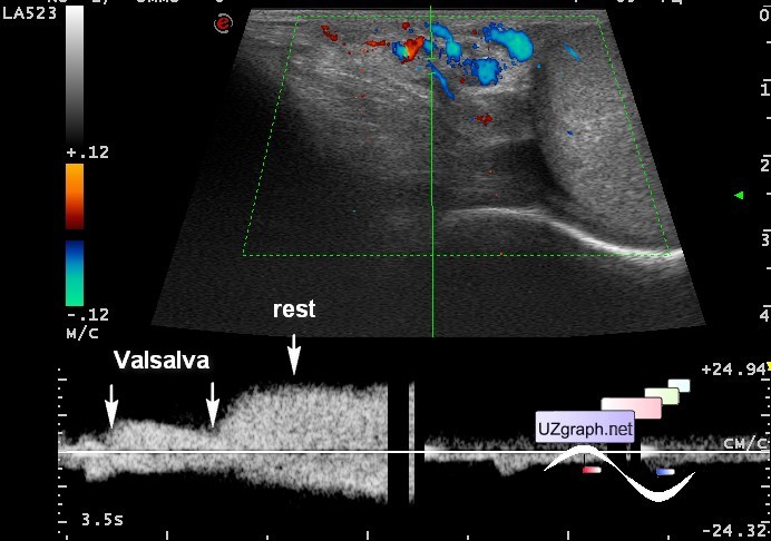
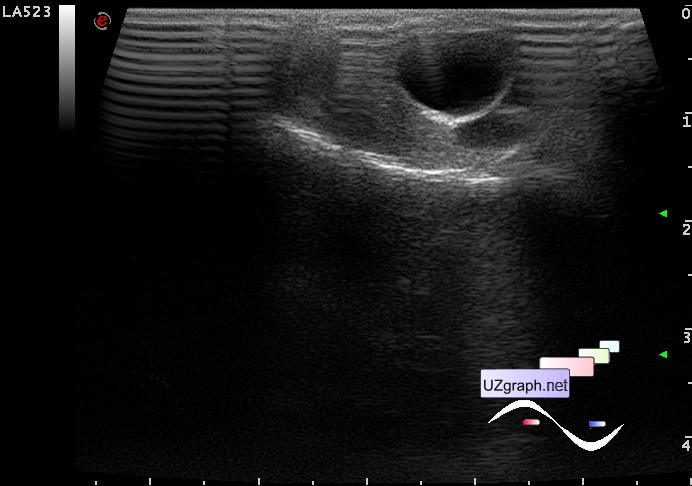
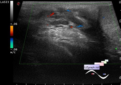
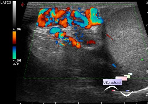
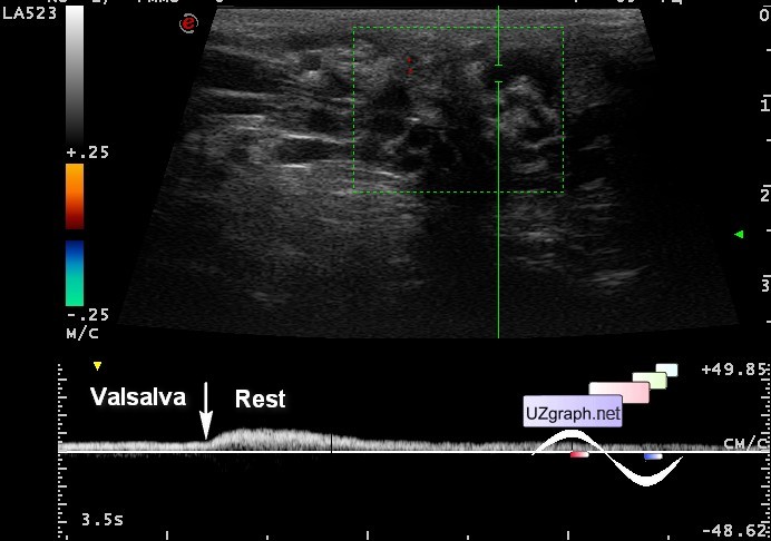
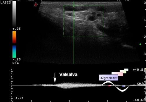
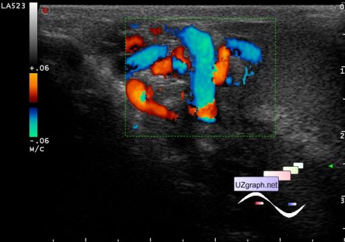
Teenager coming for re-ultrasonography with diagnos of varicocele on the left, in the previous ultrasound there is a doubtful Valsalva maneuver.
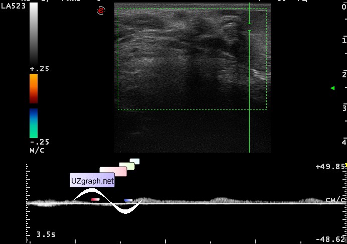
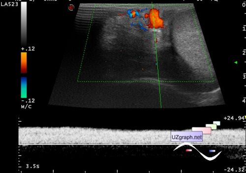
In the current US on CFM: veins of the pampiniform plexus: lying upto 1 mm, Valsalva maneuver negative; standing upto 4 mm, Valsalva maneuver also negative. In the spectral doppler(PW) standing Valsalva maneuver negative, sometimes paradoxical - in response to a maneuver there is a decrease of bloodflow velocity. Vmax standing upto 19 cm / sec.
And a cyst in the projection of head of left epididymis ...
Files:
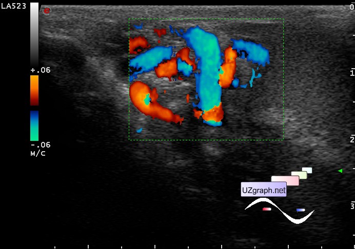
1 - CFM lying, at maneuver nothing has changed.
2 - CFM standing, at maneuver nothing has changed.
3 - PW standing
4 - PW Valsalva maneuver, by arrows indicated transitional zone - after a teenager relaxed the bloodflow velocity increased - a paradox, usually everything is exactly the opposite.
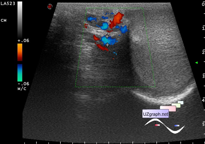
Teenager after consulting of an urologist with a clinical diagnosis of varicocele is directed to ultrasound.
On the CFM in the supine position left veins of the pampiniform plexus upto 3.5 mm, in PW visualized intermediate retrograde flow or reflux of intermediate type (1-2 seconds by Cornud et al.1999); in standing position veins upto 4 mm, in PW there is a paradoxical Valsalva maneuver, i.e. no retrograde blood flow but decrease of the blood flow velocity while Valsalva maneuver.
And relevant issue - Is PW give us anything in determining of varicocele? Is it changes our diagnosis? My answer is - no.