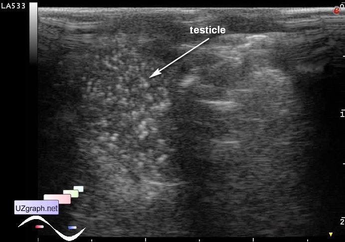
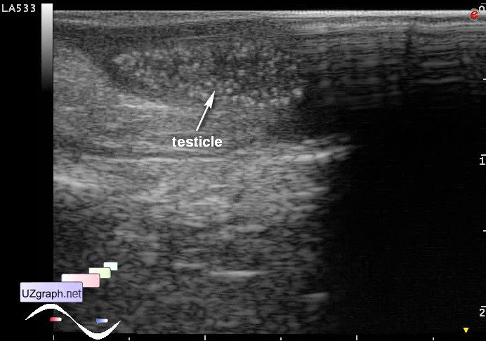
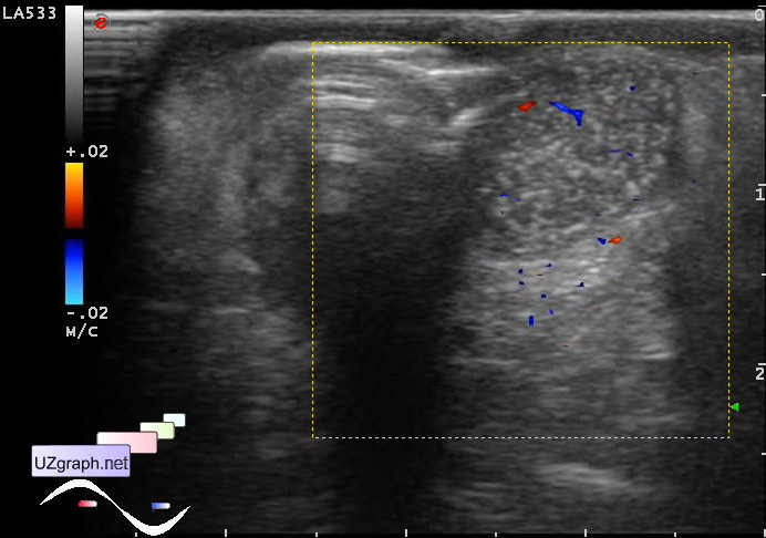
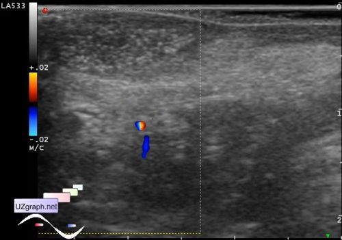
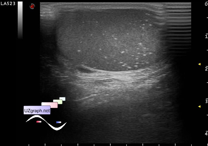
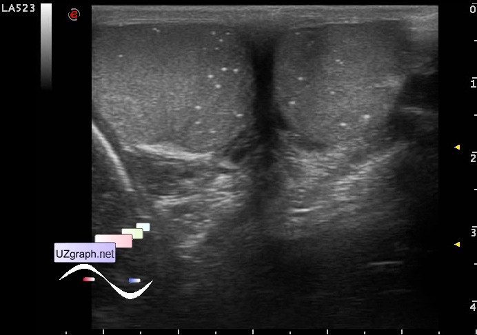
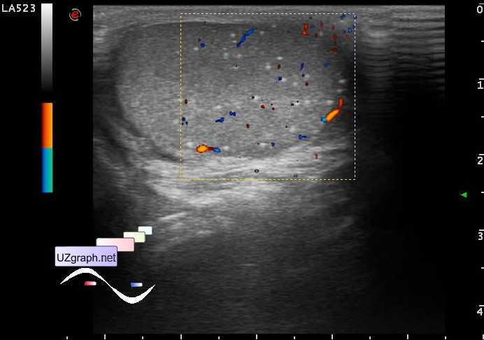
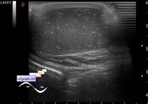
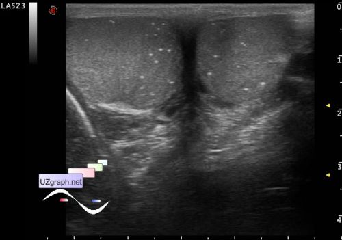
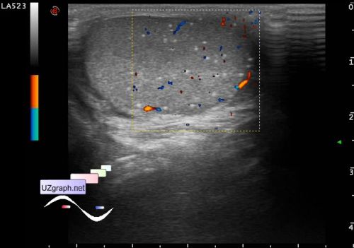
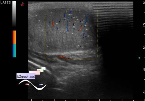
| "... testicular microlithiasis (TM)... Classic TM is arbitrarily defined by the presence of five or more microliths on at least one ultrasound image, whereas limited TM is defined by the presence of fewer than five microliths on all images; however, a large number of varying definitions have been used in the extensive sonographic literature on this topic. ... testicular cancer (TC) ... At least 20 conditions have been reported in association with TM. Other than the association with TC, which is relevant to the discussion in the present study, the more frequently reported associations include infertility, testicular atrophy, cryptorchid testicle, pulmonary alveolar microlithiasis, hypogonadism, Kleinfelter syndrome, Down syndrome, fragile X syndrome, testicular or appendiceal torsion, postorchiopexy testis, male hermaphroditism, neurofibromatosis, AIDS, and other conditions. The truly interesting question is whether these documented associations are coincidental or causal. ... Testicular microlithiasis is present without intratesticular mass or other worrisome findings. In the absence of any other risk factors for testicular cancer (e.g., personal history of testicular cancer, a father or brother with testicular cancer, history of cryptorchidism or maldescent, testicular atrophy, or other risk factors), no further imaging or biochemical follow-up is necessary; all that is recommended is routine monthly testicular self-examination. However, if the patient has risk factors for testicular cancer, referral to a urologist for evaluation and determination of an optimal follow-up strategy is recommended." external link | |