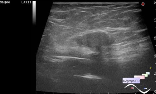
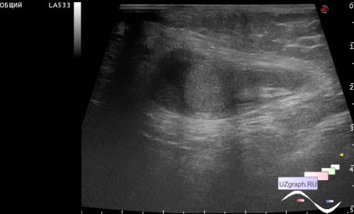
A 5-year-old child came to the emergency department of the Children City Clinical Hospital with a diagnosis of edematous scrotum syndrome on one side, and was urgently sent for an ultrasound scan.
Visually, the scrotum is empty, one half of the scrotum is pink.
On ultrasound, both testicles are reduced in size (up to 1 cm in length), one is stably located in the proximal third of the inguinal canal, the second migrates between the scrotum and the distal third of the inguinal canal, round in shape, around up to 1 ml of free fluid, the blood flow at CFM is slightly increased, next to the testicle there is a small structure (diff. diagnosis: epididymis, hydatida).