Teenager with chronic moderate pain in the scrotum was addressed to ultrasound with suspicion of hydatide torsion. In the contralateral scrotum part visualized two echogenic foci, one of them moveable.
In the literature they called scrotum pearls(stones) because optically they looks like pearls - white and rounded.
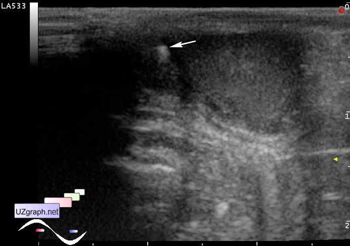
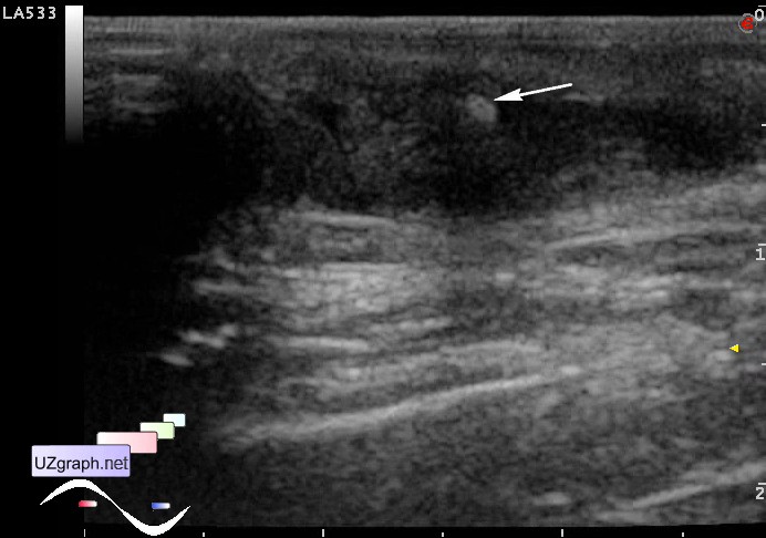
Schoolboy with complaints of pain in the left hemiscrotum is aimed to ultrasound.
At ultrasound in the left hemiscrotum slight increase of the epididymis. In the right hemiscrotum visualized hyperechoic microlesion - microstone(arrow at images).
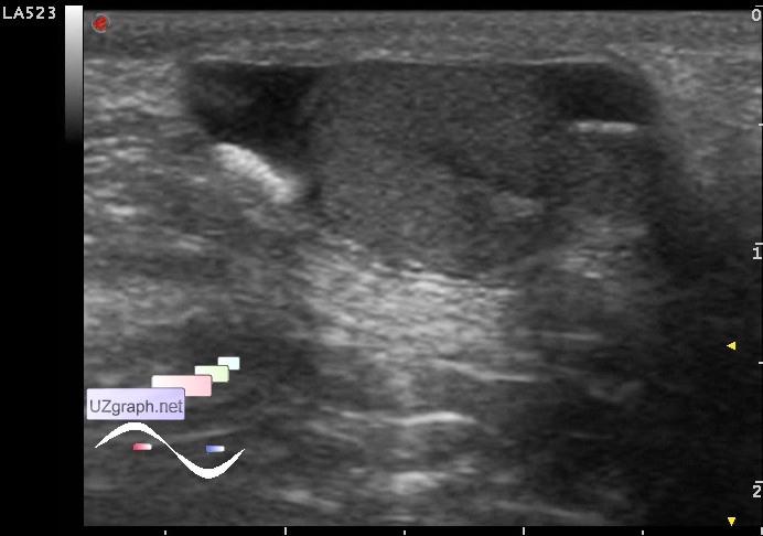
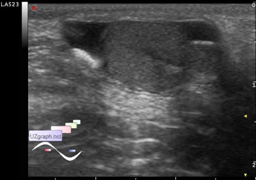
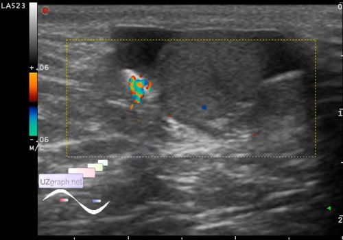
Teenager after consultation of an urologist aimed to re-ultrasonography, in the previous ultrasound (another sonologist) was suspected a cyst of some hemiscrotum.
In the current US cyst isn't visualized, there is a small hydrocele (up to 1 ml of free fluid) and a linear hyperechoic lesion about 4 x 2 mm, on the CFM gives a twinkling artifact.