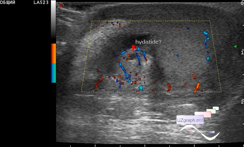
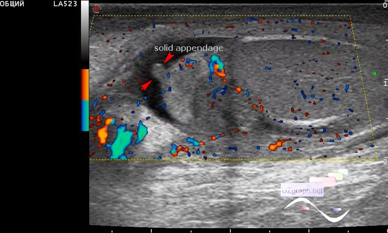
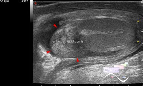
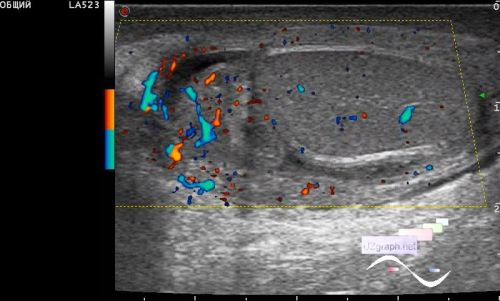
A 7-year-old child was admitted to the emergency department of a children's hospital with complaints of an enlargement and pain in the scrotum, examined by a surgeon, sent for an ultrasound scan. On ultrasound in the scrotum on the side of complaints there is an increase in the size of the epididymis, the structure of the epididymis is diffusely heterogeneous, the blood flow at CFM(DPD) is enhanced (epididymitis). The size of testicle is not enlarged, the structure is unremarkable but there is also an increase in blood flow (orchitis) at DPD. Also in the projection of the head of the epididymis is visualized single anechoic round shape lesion without blood flow at CFM(hydatide? hydatide torsion?), next to it, another appendage of the solid echostructure is visualized. Acute orchoepididymitis was suspected secondary to torsion of the Morgagni's hydatide and was recommended the second consultation of the surgeon of the emergency department. external link |