Teenager with acute pain in the scrotum day ago and following scrotal edema was addressed to ultrasound with suspected hydatide torsion.
Optically scrotum asymmetrical, appropriate side enlarged and pink color.
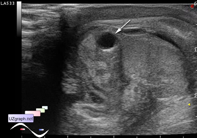
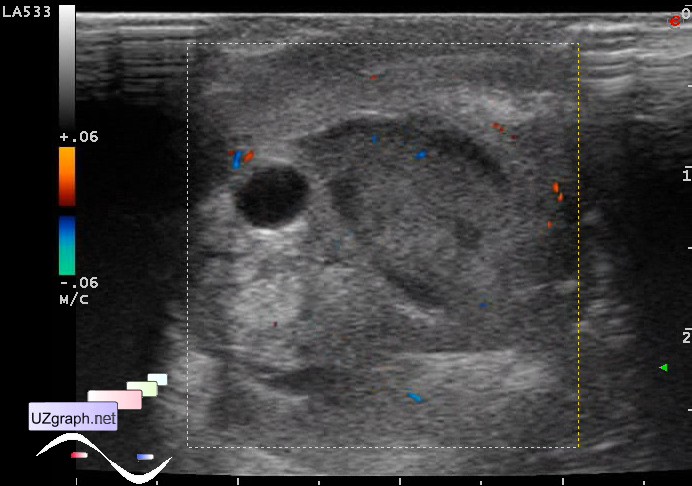
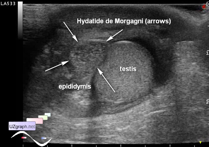
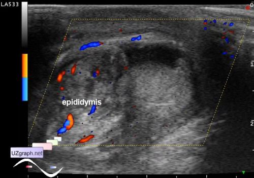
At sonography scrotum wall of appropriate side thickened, head of epididymis enlarged, with cyst inside(possible hydatide de Morgagni ultrasound appearance) and an encapsulated fluid near(possible pyocele), bloodflow in the appropriate testicle diffusely increased on CFM.
Possible diagnosis: Hydatide torsion, orchiepididymitis, pyocele.
Teenager with unilateral scrotal edema syndrome is aimed at ultrasound.
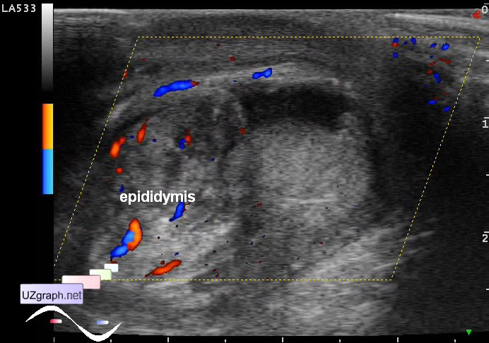
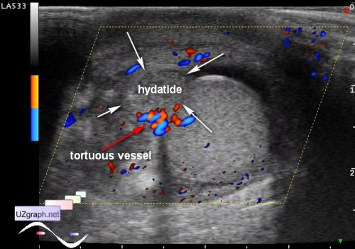
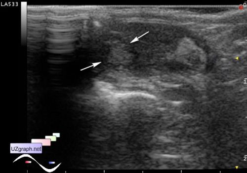
At ultrasound epididymis increased in size, has heterogeneous structure, with diffusely enhanced blood flow at DPD(file 1). In the upper third of the anterior scrotum area near epididymis visualized rounded solid mass up to 1 cm(file 2), without blood flow at DPD, posterior to this mass there is a tortuous vessel(file 3), presumably hydatide de Morgagni torsion.
PS. Hydatide de Morgagni(appendage of testis) can have different appearances: solid (hyper- or hypoechoic) or cyst.